Plotting Cranial and Spinal Nerve Pathways in a Human Anatomy Lab
Published online:
Abstract
Hands-on experiences with cadavers, animal dissections, or plastic models are effective at improving student learning and attitudes in human anatomy courses. The hands-on experience allows students to visualize and experiment with anatomical structures in three-dimensions, which is critical to learning the spatial organization of the human body. One area of human anatomy that is especially essential to understand in three dimensions is the pathways that cranial and spinal nerves take throughout the body. While human anatomy textbooks give two-dimensional and cadaver images of the routes that these nerves take, it is sometimes difficult to visualize the actual path that nerves take through the body without using a physical model. In this lesson, I describe an easily adaptable and expandable framework for students to learn about cranial and spinal nerve pathways in a human anatomy lab. Students use string or twine to model the pathways of nerves on human skeleton models and thus are able to better learn the origins, pathways, and innervation points of these nerves. This lesson will be of value to instructors of human anatomy labs who want to give their students a hands-on activity in the identification, plotting, and analysis of cranial and spinal nerve pathways.
Citation
Shaffer, J. F. 2014. Plotting Cranial and Spinal Nerve Pathways in a Human Anatomy Lab. CourseSource. https://doi.org/10.24918/cs.2014.9Society Learning Goals
Science Process Skills
- Modeling/ Developing and Using Models
- Build and evaluate models of biological systems
- Communication and Collaboration
- Share ideas, data, and findings with others clearly and accurately
Lesson Learning Goals
- Understand the structure and function of cranial and spinal nerves
- Understand how nerves travel throughout the body
Lesson Learning Objectives
- Identify and describe the functions of cranial and spinal nerves
- Identify cranial and spinal nerve origination points and what structures they innervate
- Trace the routes that cranial and spinal nerves take throughout the body
Article Context
Course
Article Type
Course Level
Bloom's Cognitive Level
Vision and Change Core Competencies
Vision and Change Core Concepts
Class Type
Class Size
Audience
Lesson Length
Pedagogical Approaches
Principles of How People Learn
Assessment Type
INTRODUCTION
Learning human anatomy requires being able to think in three-dimensions and spatially visualize the structures of the human body. One of the best ways to do this is through working with models, whether they are cadavers, animal dissections, plastic models, or computerized models. Many studies have assessed the use of anatomical models in anatomy and physiology courses, and the consensus results suggest that models are clearly beneficial to student learning and attitudes (1-6). A recent study compared the efficacy of using animal dissections, plastic models, and computer models and found that students using plastic models performed the highest on a common assessment, but students using animal dissections perceived science as more “fun” than the other groups (4). The majority of students enrolled in an undergraduate human anatomy course that used plastic models reported that the models contributed the most to their learning of the course material (5). No matter the situation or the level, the use of models in anatomy courses will have a positive effect of students.
While students may struggle to learn many anatomical systems and regions, one of the most, if not the most, problematic systems is the nervous system. Kramer and Soley surveyed South African medical students and found that neuroanatomy was the most difficult histology topic to learn and the second most difficult gross anatomy topic (second to the pelvis) (7). British dental students indicated that neuroanatomy was the most difficult subject (more difficult than histology, topographical anatomy, and embryology) (8). Anecdotally, my undergraduate students have told me that the exam covering the nervous system is always the most challenging exam of my human anatomy course.
In order to assist students with learning the nervous system, I developed a hands-on lesson for an undergraduate human anatomy course that requires students to use and create models of cranial and spinal nerve pathways. Using skeleton models and twine or string, students identify specific cranial or spinal nerves, attach them to their origination points on a skeleton model, and model their pathways through the body. Students model two cranial nerves (optic (II) and vagus (X)) as well as four spinal nerves, one from each of the four plexuses (the phrenic nerve from the cervical plexus; the ulnar nerve from the brachial plexus; the obturator nerve from the lumbar plexus; and the tibial nerve from the sacral plexus). These nerves were chosen because they are commonly studied in undergraduate human anatomy courses and they also exhibit a wide range of features (origination point, innervation point, length, etc) that students can model in this lesson. Students should be familiar with the basic structure and function or and terminology related to cranial and spinal nerves before completing this lesson.
The main form of active learning used in this lesson is collaborative learning in which students work together to solve a set of problems, with the goal of enhancing what they learn about the topic (9). Collaborative learning has been used in a variety of human anatomy and physiology courses with positive affects. Undergraduate human physiology laboratory courses have used collaborative learning and active learning to improve student performance on respiratory physiology modules (10). Medical school gross anatomy laboratory courses that used collaborative learning strategies saw increased exam performance (11) and positive students attitudes towards the instructional methodology (11, 12). Additionally, a multi-year study of the “Teams and Streams” model showed that incorporating collaborative learning and inquiry-based labs into undergraduate biology laboratory curricula improved student performance on exams and were favorably evaluated compared to students who had traditional laboratories (13).
This lesson addresses key learning objectives related to the nervous system including “identify and describe the functions of cranial and spinal nerves,” “identify cranial and spinal nerve origination points and what structures they innervate,” and “trace the routes that cranial and spinal nerves take throughout the body.” Student evaluation data suggests that this is an effective and enjoyable lesson to teach students about cranial and spinal nerves in a human anatomy lab.
Intended audience
The lesson was designed an upper-division human anatomy course that taught human anatomy using a systemic approach. The students were a mix of junior and senior biological sciences majors and sophomore nursing science majors. This course has both a lecture and a lab component and required a human physiology lecture course as a pre-requisite. The lab sections where this lesson was implemented met in groups of 24 students. This lesson is appropriate for students majoring in the biological, nursing, or other health sciences. Additionally, this lesson could be appropriate for advanced high school students who are taking anatomy and physiology courses at the high school level.
Learning time
This lesson is part of a larger three-hour lab session on the nervous system. The lesson takes about 45 minutes to complete. If a lecture component is included before the lesson, then the required time will be increased to ~60 minutes. See Table 1 for a breakdown of the time required.
Pre-requisite knowledge
Students should be able to perform the following objectives before attempting this lesson:
- Describe the basic gross anatomical structure of a nerve
- Explain how cranial and spinal nerves originate from the brain and spinal cord, respectively
- Identify cranial and spinal nerves in photographs or on models
- Describe what a nerve plexus is and how spinal nerves enter and exit plexuses
- List the major spinal nerves that are associated with each nerve plexus
Students can obtain this knowledge via the lecture part of the class, through pre-class homeworks or assignments (either original or those from online learning systems such as Mastering A&P or Connect), or from a lecture in lab just prior to the lesson see Supplemental File S6 for an outline of a sample lesson). Students are not expected to know the detailed pathways of the nerve priors to this lesson (i.e. following the anterior or posterior side of a limb) as they will model these pathways in the lesson.
SCIENTIFIC TEACHING THEMES
Active Learning
Students will actively engage in learning the concepts by working in groups to accurately trace the paths of nerves throughout the body. Students will be given an envelope with six unlabeled “nerves” and based on what they know about nerves they will have to identify each one and then attach them to a skeleton model correctly. The entire lesson is conducted by students in small groups of 3 to 5.
Assessment
Measurement of learning takes place in two ways in this lesson. First, the instructor will conduct formative assessment by checking each group’s skeleton models after they have attached their nerves. At this point, the instructor can ask for clarification about why a certain nerve is following a certain path, and also to ask students to identify the nerves that they modeled. At the end of the lesson there is a summative assessment in the form of a worksheet that the students work on as a group and turn in for credit. Answer keys for the student handout (Supplemental File S1) and the group worksheet (Supplemental File S3) are included in supplementary materials (Supplemental File S2 and S4, respectively).
Inclusive Teaching
Students work in small groups of 3 to 5 for the entire lesson, so they are learning to work with diverse students and how to handle group work environments. This lab also requires them to translate two-dimensional visual information to a three-dimensional model, thus requiring students to use multiple modalities and points of view.
LESSON PLAN
The following lesson plan contains three parts: materials required, instructor preparation, and the lesson description. The preparation takes about one hour to complete and the lesson takes about 45 minutes to complete. Please refer to the Teaching Discussion section for suggestions of other possible materials that you can use for this lesson. Table 1 depicts the timeline of the entire lesson.

Tables 1, 2 and 3. Plotting Nerve Pathways
Materials required
- Human skeleton models (life-size models are best, but smaller ones would work too; we use the following model: http://www.a3bs.com/super-skeleton-model-sam-a13,p_164_15.html)
- Human brain model (one that fits into the skull of the skeleton; we use the brain from the following torso model: http://www.a3bs.com/classic-unisex-torso-14-part-b13,p_58_192.html)
- Human eyeball model (one that fits into the orbital socket of the skeleton: we use the eyeball from the following torso model: http://www.a3bs.com/classic-unisex-torso-14-part-b13,p_58_192.html)
- String / rope / twine – try to find a product that does not fray or unwind easily
- Tape (masking or lab tape is best)
- Measuring tape / ruler
- Scissors
- Envelopes
Instructor preparation
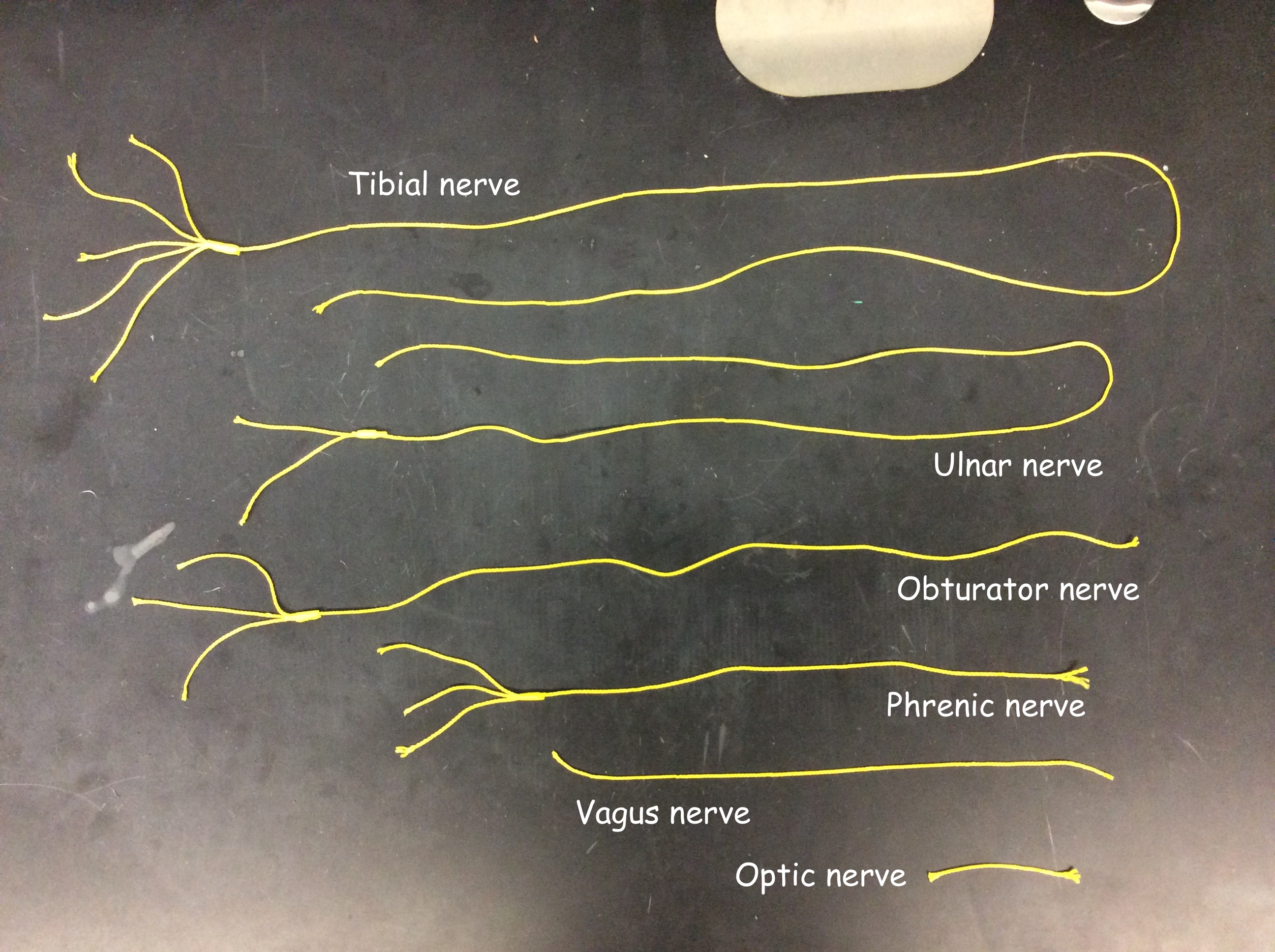
For the first time through this lesson, you will need to make the six “nerves” from the string. Each group of students will need one set of six nerves, so you will need to repeat the following process for however many groups you will have. Depending on how carefully students use the nerves, you may need to make new ones every time, or fix them as needed.
- First you will need to make the nerves using the string. Table 2 lists the lengths of string and number of spinal nerve attachment points to be used for the nerves emerging from nerve plexuses.
- To make a nerve, cut the required length of string from Table 2, and then attach the number of spinal nerve attachment points to one end using tape. NOTE: These lengths are approximate and work well for the specific model of skeleton that I used to develop this lesson. If you have a different-sized model, you will want to adjust the lengths of your nerves as needed.
- Each spinal nerve attachment point can be a 3 to 4” piece of string (pieces of pipe cleaners can be used as well and may be more effective). The spinal nerve attachment points will be taped or otherwise attached to the spinal nerves that emerge from the skeleton model (or onto the neighboring vertebrae if the model does not have spinal nerves), so these are the parts that will be subjected to the most damage from students. Figure 1 shows examples of the completed set of six nerves. As described in the Teaching Discussion section, the instructor can choose to use different colored strings to represent different nerves or other materials (pipe cleaners, Velcro-backed adhesive, Bendaroo, etc) to make the nerve attachment points.
- Place one set of six nerves into an envelope. Each group completing the lesson will be given one envelope.
- Repeat steps 2 to 4 for however many groups you have in your class.

Figure 1. Completed set of six nerves used in this lesson.
Lesson description
If students have not been exposed to cranial and spinal nerves yet, the instructor may wish to give a 15-minute lecture on the basic structure and function of cranial and spinal nerves. Please see Supplemental File S6 for an outline of a possible optional lecture.
Have students get into groups of 3 to 5. Each student should have a copy of the lesson handout (Supplemental File S1). Give each group the following:
- One envelope containing the “nerves” you prepared ahead of time
- A human skeleton model
- A midsagittal-model of the human brain that fits inside the skeleton’s skull
- An eyeball model that fits in the orbital socket of the skeleton
- A roll of tape (lab tape or masking tape works best)
- One group worksheet (Supplemental File S3) – to be completed after the lesson is finished
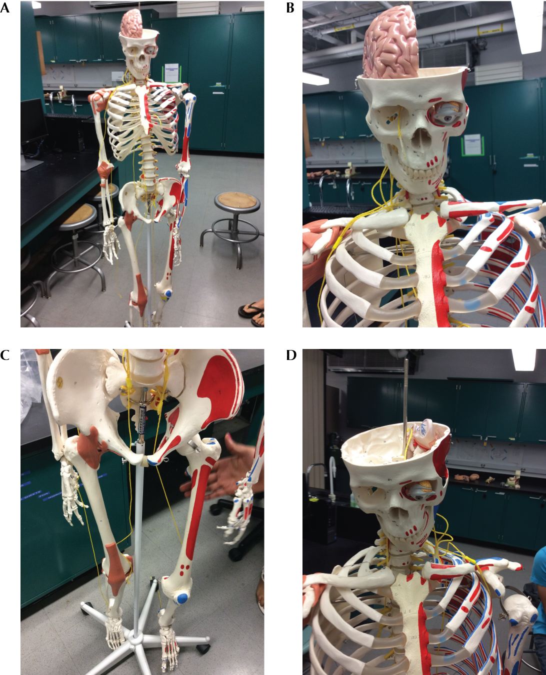
Allow students to work on the lesson handout (Supplemental File S1) as a group. The lesson handout contains detailed instructions for what the students are to do, which the instructor should tell each group to read carefully before beginning. However, the instructor may want to emphasize that there are six different nerves, and that they need to identify the nerves before attempting to attach them to the skeleton. The instructor may want to check on each group at this point, as sometimes students may have difficulties in identifying the nerves. As there usually is one instructor per 24 students (in six groups of four students), the instructor may wish to spend two minutes or so with each group as needed and then rotate through the groups to make sure they are on track and to answer any questions. Common student questions include locating the correct skull foramina and whether or not they should be exact in modeling the nerve pathways (the answer is yes, as much as possible!). The groups will call the instructor over at each checkpoint in the lesson to assess their progress on the nerve pathways. Common student errors include passing nerves in the wrong location relative to a bone (e.g. anterior to the femur instead of posterior) and choosing wrong spinal nerve attachment points.Supplemental File S2 includes the answers for the questions in the student handout (Supplemental File S1). Figures 2A to 2D show photographs of sample student nerve pathways.

Figure 2. Photographs of sample student nerve pathways.
Once each group is complete, have them complete the group worksheet (Supplemental File S3) which is to be turned in for credit. Supplemental File S4 is the answer key for this worksheet.
When each group is finished, they should take a photograph of their completed models and then carefully remove each of their nerves and place them back in the envelope.
TEACHING DISCUSSION
The main goal of this lesson was to give students an opportunity to model the pathways of nerves through the body, from origination point to innervation target. Four different instructors in six lab sections have implemented this lesson over two quarters at UC Irvine (Spring and Summer Session II, 2014) for a total of 135 students (~24 students per lab section). The fact that four instructors have implemented this lesson multiple times provides evidence that this lesson is transferable and readily adopted by new instructors. The lesson is largely student-driven, as once the students are given the lesson handout (Supplemental File S1) they can read the detailed instructions and proceed on their own in their groups. The instructor should circulate constantly to make sure all groups are on task and to answer any questions that may arise. When students were working on this lesson, they were fully engaged, first to figure out which nerve is which, and then to attach and trace the path of the nerve on the skeleton model. Students were working actively in groups to perform these tasks, with one student counting off the spinal nerves, one attaching the nerves to the model, and another taping the nerves in place as they passed over the model. The students also seemed to be having a lot of fun with this lesson, especially when removing the brain and eyeball and placing them into the skeleton model. They also enjoyed taking pictures of their completed models at the end of the lesson.
Student evaluations
Students enrolled in the Applied Human Anatomy course at UC Irvine in Summer Session II (2014) completed this activity as part of their normal lab on the nervous system and evaluated the activity after its completion. Students were asked to evaluate the lesson by marking their level of agreement with six Likert-type statements as well as an open-ended question asking for improvements in the lesson. The results are summarized in Table 3 below (n = 39). Overall the students rated the lesson highly, especially for being able to achieve the learning outcomes related to nerve pathways and function. The students also rated the lesson highly in terms of it being fun and also challenging. The most frequent open-ended responses from students included comments about minor difficulty in attaching the nerve attachment points to the skeleton models. A suggestion for how to improve this potential issue is provided below.
Possible modifications / adaptations
This lesson can be easily modified, adapted, and expanded to suit the needs of any instructor. Below are some possibilities for modifications.
- Additional / different nerves – Instead of making the nerves listed in Table 2, an instructor can choose whatever cranial and spinal nerves that they want the students to model
- Students design the nerves – Instead of making the nerves yourself ahead of time, you can have the students make their own nerves by cutting the string and affixing the spinal nerve attachment points. This will require more time so plan accordingly. You can also have different groups make different nerves, and then have them attach them all to a common skeleton to show the cumulative efforts of the class in modeling nerve pathways through the body.
- Different materials – If you cannot or do not want to use string, twine, or mason’s line, you can use other types of materials to make the nerves. Pipe cleaners, Velcro-backed adhesive, or Bendaroos may be more effective for the nerve attachment points. Some frequent student comments included that the materials were not durable and that it was difficult to tape the nerves onto the skeleton, so using other types of materials may be warranted. An additional possibility would be to make the nerves from different colored string or twine to more easily visualize the different nerves on the skeleton model.
SUPPLEMENTAL MATERIALS
Supplemental File S1. Plotting Nerve Pathway – Lesson handout
Supplemental File S2. Plotting Nerve Pathway – Lesson handout key
Supplemental File S3. Plotting Nerve Pathway – Group worksheet
Supplemental File S4. Plotting Nerve Pathway – Group worksheet key
Supplemental File S5. Plotting Nerve Pathway – Student evaluation form
Supplemental File S6. Plotting Nerve Pathway – Sample optional lecture on cranial and spinal nerves
ACKNOWLEDGMENTS
I would like to thank the students of Bio Sci D170 who have (hopefully!) benefited from this lesson. I would also like to thank Pavan Kadandale and Brian Sato and the reviewers for helpful comments that have improved this lesson.
References
- DeHoff ME, Clark KL, Meganathan K. 2011. Learning outcomes and student-perceived value of clay modeling and cat dissection in undergraduate human anatomy and physiology. Adv Physiol Educ 35:68-75.
- Haspel C, Motoike HK, Lenchner E. 2014. The implementation of clay modeling and rat dissection into the human anatomy and physiology curriculum of a large urban community college. Anatomical sciences education 7:38-46.
- Waters JR, Van Meter P, Perrotti W, Drogo S, Cyr RJ. 2005. Cat dissection vs. sculpting human structures in clay: an analysis of two approaches to undergraduate human anatomy laboratory education. Adv Physiol Educ 29:27-34.
- Lombardi SA, Hicks RE, Thompson KV, Marbach-Ad G. 2014. Are all hands-on activities equally effective? Effect of using plastic models, organ dissections, and virtual dissections on student learning and perceptions. Adv Physiol Educ 38:80-86.
- Wright SJ. 2012. Student perceptions of an upper-level, undergraduate human anatomy laboratory course without cadavers. Anatomical sciences education 5:146-157.
- Lujan HL, Krishnan S, O'Sullivan DJ, Hermiz DJ, Janbaih H, DiCarlo SE. 2013. Student construction of anatomic models for learning complex, seldom seen structures. Adv Physiol Educ 37:440-441.
- Kramer B, Soley JT. 2002. Medical students perception of problem topics in anatomy. East African medical journal 79:408-414.
- Parkin IG, Rutherford RJ. 1990. Feedback from dental students: performance in an anatomy department. Medical education 24:27-31.
- Bruffee, K. 1993. Collaborative Learning. Baltimore, MD: The Johns Hopkins University Press.
- Modell HI, Michael JA, Adamson T, Horwitz B. 2004. Enhancing active learning in the student laboratory. Adv Physiol Educ 28:107-111.
- Vasan, NS, DeFoww D. 2005. Team learning in a medical gross anatomy course. Medical Education 39:524.
- Krych AJ, March CN, Bryan RE, Peake BJ, Pawlina W, Carmichael SW. 2005. Reciprocal peer teaching: students teaching students in the gross anatomy lab. Clinical Anatomy. 18:296-301.
- Luckie DB, Maleszewski JJ, Loznak SD, Krha M. 2004. Infusion of collaborative inquiry throughout a biology curriculum increases student learned: a four-year study of "Teams and Streams." Adv Physiol Educ 287:199-209.
Article Files
Login to access supporting documents
Plotting Cranial and Spinal Nerve Pathways in a Human Anatomy Lab(PDF | 756 KB)
Supplemental File S1. Plotting cranial and spinal nerve pathways in a human anatomy lab-.docx(DOCX | 135 KB)
Supplemental File S2. Plotting cranial and spinal nerve pathways in a human anatomy lab-.docx(DOCX | 147 KB)
Supplemental File S3. Plotting cranial and spinal nerve pathways in a human anatomy lab-.docx(DOCX | 63 KB)
Supplemental File S4. Plotting Nerve Pathway Group worksheet key.docx(DOCX | 16 KB)
Supplemental File S5. Plotting cranial and spinal nerve pathways in a human anatomy lab-.docx(DOCX | 67 KB)
Supplemental File S6. Plotting cranial and spinal nerve pathways in a human anatomy lab-.docx(DOCX | 119 KB)
- License terms

Comments
Comments
There are no comments on this resource.