Building a Model of Tumorigenesis: A small group activity for a cancer biology/cell biology course
Published online:
Abstract
The multistep nature of tumorigenesis is a foundational concept in the context of Cancer Biology. Many students do not appreciate the complex nature of cancer development nor do they understand how scientists are able to unravel the molecular pathways that lead to tumorigenesis. In this small group activity, students are presented with background information about the multistep nature of tumorigenesis and complete a priming activity that allows them to brainstorm and discuss experimental design. Students are then presented with data from the landmark manuscript, published in 1998 by Vogelstein et al., describing the first pathway of genetic alterations associated with colorectal tumor development. Using selected pieces of the manuscript, students answer discussion questions and analyze the data presented in the paper. Using their analysis, students are able to create a scientifically valid molecular model of colorectal development that matches the model presented in the literature. The group activity can be followed by a whole class discussion about current knowledge about colorectal tumor development.
Citation
Wright, L.K. 2015. Building a Model of Tumorigenesis: A small group activity for a cancer biology/cell biology course. CourseSource. https://doi.org/10.24918/cs.2015.18Society Learning Goals
Cell Biology
- Cell cycle and cell division
- How do cells conduct, coordinate, and regulate nuclear and cell division?
Genetics
- Genetic variation
- How do different types of mutations affect genes and the corresponding mRNAs and proteins?
Science Process Skills
- Process of Science
- Locate, interpret, and evaluate scientific information and primary literature
- Interpret, evaluate, and draw conclusions from data
- Construct explanations and make evidence-based arguments about the natural world
- Modeling/ Developing and Using Models
- Build and evaluate models of biological systems
- Communication and Collaboration
- Share ideas, data, and findings with others clearly and accurately
Lesson Learning Goals
- Students will appreciate the complex nature of human tumorigenesis.
- Students will understand the importance of retrospective clinical studies.
- Students will appreciate the importance of modeling.
Lesson Learning Objectives
At the end of the activity, students will be able to:- Analyze data from a retrospective clinical study uncovering genetic alterations in colorectal cancer.
- Draw conclusions about human tumorigenesis using data from a retrospective clinical study.
- Present scientific data in an appropriate and accurate way.
- Discuss why modeling is an important practice of science.
- Create a simple model of the genetic changes associated with a particular human cancer.
Article Context
Course
Article Type
Course Level
Bloom's Cognitive Level
Vision and Change Core Competencies
Vision and Change Core Concepts
Class Type
Class Size
Audience
Lesson Length
Pedagogical Approaches
Principles of How People Learn
Assessment Type
INTRODUCTION
Human tumorigenesis is a complex, multistep process involving numerous molecular alterations that develop over time (1-5). Novice biology students may think that cancer develops "all at once" when, in reality, precancerous cells undergo a number of genetic changes over time. These genetic and/or epigenetic changes enable precancerous cells to acquire phenotypic changes that allow them to eventually become a malignant growth. The time-span between the earliest initiating genetic/epigenetic event and the time of cancer diagnosis ranges however, in some cases, it may be decades.
Human cancer is an incredibly complex set of diseases. Typical undergraduate students may wonder how research scientists or other biology "experts" can begin to understand and ask questions about a disease like cancer. The activity presented here not only allows students to think and reason about the multistep nature of human cancer using a landmark paper (6), it also allows students to construct a scientific model to helps explain the multi-step nature of this disease. Modeling is an important scientific practice that spans STEM disciplines and is called out in many reform initiatives in K-12 and higher STEM education (7,8). Models help experts explain various scientific phenomena, formulate research questions, and design experiments. Providing undergraduate students with opportunities to practice scientific modeling may even begin to help them develop more expert-like behaviors.
Most instructors would agree that reading primary scientific literature is important for the development of scientifically literate biology majors. Considering that moving students toward expert-level thinking and reasoning is an overarching goal of undergraduate science education (7,8,9,10), activities that involve analysis and discussion of the primary scientific literature should be brought into the classroom. Most instructors would also agree, though, that facilitating these activities in a classroom setting could present a number of challenges. For example, many students may lack the skills and/or confidence to successfully navigate a manuscript from the primary literature. Without appropriate scaffolding and instructor support, students often get bogged down by complex scientific jargon, are miffed by the methodology section, and more-or-less give up by the results and discussion sections. In addition to student preparation issues, facilitating a whole-class discussion of the literature can be challenging from the instructor point of view. How does an instructor manage a whole-class discussion with a large number of students? How does one encourage everyone in the class to participate when only a minority of students actually managed to get through the entire paper?
In reality, even biology experts such as us read the scientific literature for a variety of reasons. Sometimes, we are interested in just the methodology section so we can recreate a protocol in our own laboratories. Other times, we scan the introductory sections and just focus on the results and, of course, some papers demand a very deep reading and analysis. The activity presented here takes the approach of having students read just enough of a scientific manuscript to gain a basic understanding of the research study. The activity allows students to analyze data from a retrospective clinical study, present scientific data in an appropriate and accurate way, and create a simple model of the genetic changes associated with colorectal cancer by reading, analyzing, and discussing portions of a scientific paper. This strategy is manageable for many different classroom settings and allows all students to participate in discussion of the primary literature in a guided and organized way.
This lesson incorporates a priming activity, which is a strategy that can promote deep learning. Education research has shown that allowing students to make predictions and invent models or formulas before a "correct answer" is discussed can be an effective way to improve student learning (7,8,9,10,11,12). In this lesson, the priming activity asks students to design an experiment that would uncover the molecular alterations associated with progression of a particular cancer. This complex scenario allows students to think creatively and deeply about how scientists solve real-life biological problems. After groups share their ideas and discuss experimental strategies, they are primed to investigate the data presented in the activity. At the end of the lesson, nearly all students will be able to create a scientifically accurate model of tumorigenesis based on actual data. Thus, this activity may not only help students understand the complex nature of tumorigenesis and appreciate the importance of scientific models, but may also increase students' confidence in their analytical and reasoning abilities.
This activity was designed for a cancer biology course but could also be implemented in a cell biology course that covers basic aspects of cancer biology. It would be appropriate for biology majors who have completed at least one year of introductory biology, have knowledge of tumor suppressor genes and oncogenes, and are familiar with basic terminology such as "benign" and "malignant." The activity was implemented in one 75-minute class in week eleven of a fifteen-week semester course on Cancer Biology, but could easily be adapted for other settings.
SCIENTIFIC TEACHING THEMES
Active Learning
Students do a priming activity in class using a think/pair/share strategy. Small group work followed by a classroom discussion allows students to remain engaged with the material/activity for the duration of class.
Assessment
Pre-assessment: None
Post-assessments: Creation of a scientifically accurate figure based on data presented in the activity; construction of a scientifically accurate model of tumorigenesis based on the data presented in the activity.
Inclusive Teaching
The lesson involves cooperative group work and discussion and implements inclusive teaching strategies in the classroom. Since this is an activity for small groups, the instructor has a number of choices on how to sort students in the class. Allowing students to select their own groups, for example, may help them create a socially supportive environment for learning. The instructor could also pre-select groups to encourage students to interact with people they may not normally work with. This strategy could help students learn to value diverse perspectives and opinions. Because the activity does not demand expert-level knowledge about cancer biology or another sub-discipline of biology, it can be used with students with different backgrounds and abilities.
LESSON PLAN
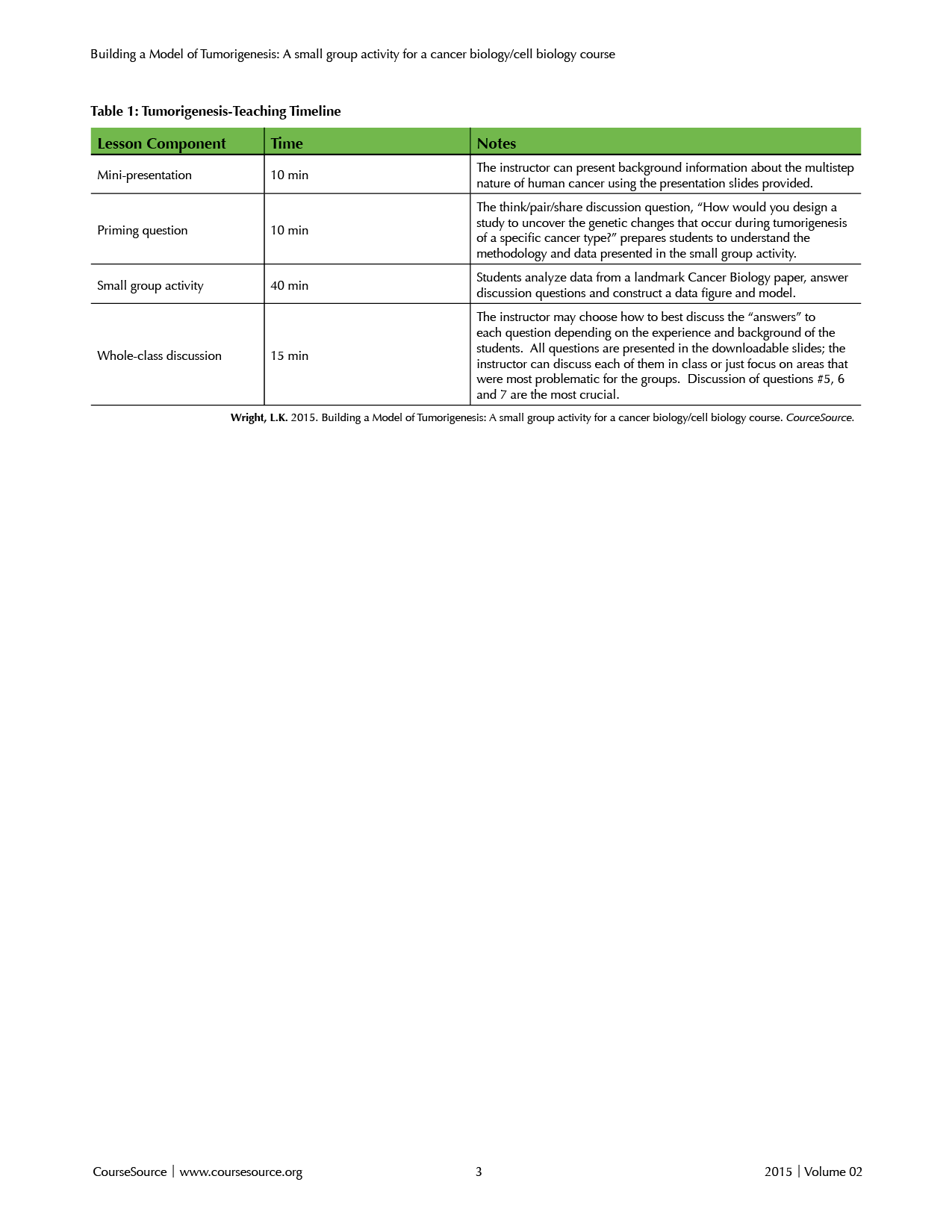
Table 1 depicts a suggested timeline for this Lesson. The mini-presentation on the multistep nature of human cancer (10 minutes) is followed by the priming activity (10 minutes). The small group activity takes the majority of class time (40 minutes) leaving 15 minutes for the final whole-class discussion. The activity was implemented in a single 75 minute period, but could easily be implemented over two class periods, especially if the introduction or discussion sections were expanded.

Table 1. Tumorigenesis-Teaching Timeline
Pre-class Preparation
Many cell biology and genetics textbooks contain at least one chapter on "Cancer," providing the instructor with many choices on how to best prepare their class for the activity. Depending on the class, the instructor may choose to assign a pre-class reading from the course textbook, such as Chapter 16 "Cancer" from Cell and Molecular Biology (13) or a chapter section entitled, "Normal Controls on Cell Division are Lost During Cancer" from Essentials of Cell Biology (15) may a useful open-access resource available through the Scitable Nature Education site (http://www.nature.com/scitable/ebooks/essentials-of-cell-biology-14749010/122997842#bookContentViewAreaDivID).
The instructor should download (and edit, if applicable) the presentation slides on the multistep nature of human cancer (Supplemental File S1. Tumorigenesis-Class Slides). Depending on instructor goals, class-time, and student background, the instructor may want to supplement the provided slides with figures of colon histology and/or images of histopathological changes associated with colorectal cancer. Neither students nor instructors are expected to have expertise in the analysis of histological samples and the activity does not depend on this knowledge. However, students may be interested in seeing differences between normal and tumor histological specimens. Note: the priming activity and slides containing images/questions from the activity are included in the presentation file. The instructor should also make enough copies of the activity (Supplemental File S2. Tumorigenesis-Class Handout) so that all students have their own copy.
In-class Actvities
Mini-presentation by Instructor
The instructor should spend the first ~10 minutes of class time presenting background information about the multistep nature of human cancer (Supplemental File S1). Tumorigenesis-Class Slides If the instructor wants to spend more than 75 minutes on this entire activity, he/she may choose to add more content to the background presentation. For example, the instructor may want to spend time reviewing the function of the protein product of the Adenomatous polyposis coli (APC) tumor suppressor gene. The point of the mini presentation is to give the students the necessary context to complete the upcoming activity.
Priming Activity
Slide #10 in the presentation illustrates a generic pathway of multistep tumorigenesis. The next slide (#11) presents the question, "What are the genetic alterations that occur at each step in the pathway?" The instructor should not answer this question but should advance to the next slide (#12) containing the priming activity: a think/pair/share discussion question that asks, "How would you design a study to uncover the genetic changes that occur during tumorigenesis of a specific cancer type?" The instructor should encourage students to think about this question individually for a few minutes and then share their ideas with their neighbors (a think/pair/share strategy). The instructor should say that ideas do not have to be limited by technology or methodology and encourage students to be creative. The instructor could walk around the room and listen to student discussion and/or probe/guide students who seem to be stuck.
After students have had time to discuss their ideas, the instructor should ask a number of different groups to share their ideas. The instructor can outline the ideas on a white-board, document camera, or computer screen projection for the class to see. It is important that the instructor not judge the likeliness of success for any group idea; the point of the priming exercise is to challenge the students to think and to allow them to grapple with a very complex scientific problem. For example, one of my groups described a scenario in which tiny video cameras are implanted in experimental mice that have been induced to develop a particular cancer; scientists may biopsy or sacrifice the experimental animals at various points along tumor development and analyze tumor specimens for genetic alterations. Other groups have proposed working with a known animal model of a particular cancer (e.g. a genetically altered mouse that is prone to developing breast cancer) and suggest sacrificing large numbers of the experimental animals and performing genomic/genetic assays at many different time points over a defined period. Although these approaches are not currently feasible for technical or ethical reasons, completing the suggested experiments could provide information about tumorigenesis. This kind of thinking should be encouraged and highlighted.
Usually at least one group will suggest using tumor samples from human patients, which is the critical point for this activity, but may struggle to figure out how to put these ideas in the context of an experimental study. In this case, the instructor might ask a few probing questions to lead students to the idea of a retrospective clinical study such as:
- "What kind of cancer would you focus on?"
- "How many samples do you think you would need?""Are you going to wait for individuals to develop this cancer and then ask to take biopsies? How long would this take?"
- "Did you know that medical institutions save/bank portions of all biopsies and medical samples as frozen or preserved tissue specimens? What if you could get access to those samples?"
When students learn that medical centers often store portions of tumor specimens, students begin to think about the possibility of using these clinical specimens in an experimental setting. Although ethical issues are not an explicit part of the lesson, an instructor may choose to expand the activity by incorporating a discussion of informed consent, protection of patients' names, and ethical conduct of research on human subjects. Once the term "retrospective study" has been introduced, the instructor should hand out the worksheets and proceed with the small group activity.
Small Group Activity
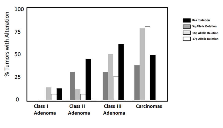
Students are given ~40 minutes to work through the activity (Supplemental File S2. Tumorigenesis- Class Handout), answer the guiding questions, and discuss ideas with their group members. The instructor should walk around the room, listen in on discussions, and answer questions when appropriate. If a particular question seems to be problematic for many of the groups, the instructor should designate time to address this question with the class. The instructor should also take note about how the student groups have chosen to present their data to question #5 in the activity, as there is more than one way to appropriately present the data. Most of the students will probably decide to construct a bar graph, as the one presented in Figure 1, but a line graph was actually used in the original publication. If students choose to present their data using a line graph, the instructor should recognize these students for thinking just like the authors of the paper. Either format works equally well.

Figure 1. Example of bar graph that students would construct using the data in the activity based on “Genetic Alternations During Colorectal-Tumor Development” by Vogelstein et al. 1988.
Whole Class Discussion
The instructor should choose how to best discuss the "answers" to each question depending on the experience and background of the students. All questions are presented in the downloadable slides (Supplemental File S1. Tumorigenesis-Class Slides); the instructor can discuss each question in class or just focus on questions that were most problematic for the groups. Discussion of questions #5, #6, and #7 is the most crucial.
When students have completed the worksheets, the instructor should ask volunteers from selected groups to draw their graphs/figures for question #5 on the whiteboard or other means (e.g. a group member could email a photo of the graph to the professor, who can then project it from his/her computer). Most groups will probably make bar graphs, but some groups may decide on a line graph. If all groups have created a bar graph, the instructor may challenge the class to think of alternate ways in which the data could be presented. Because it will take time for students to draw their graphs, encourage volunteers from all groups to also come to the whiteboard and indicate the points at which the four genetic alterations (Ras mutation, 5q loss, 18q loss, and 17p loss) occur on the model of colorectal progression.
To solve the model, students will need to recognize that certain genetic alterations are more common in different classes of tumors. Having the students create the figure/graph required for question #5 should help guide them to this realization. For example, the largest increase in tumor specimens with Ras mutations occurs between Class I and Class II adenomas. This increase strongly suggests that Ras mutations are crucial in the progression of Class I to Class II adenomas.
If instructors want to expand on the activity, they may present the slightly more complex model of colorectal tumorigenesis published in 1990 by the same group (16). Instructors may also wish to present a more current understanding of the disease as described, for example, by Walther and colleagues (17). The textbook that was used in this particular course, "The Biology of Cancer," also contains several figures related to progression of colorectal cancer (5).
TEACHING DISCUSSION
Reactions to the Lesson
Most students enjoyed the activity and expressed some degree of pride when they discovered their model agreed with the expert model. Many biology students are, unfortunately, "math phobic" and panic when asked to do even simple quantitative operations. By purposely omitting final calculated values from the data table, this activity gives students an opportunity to practice simple math in a low-stakes environment; with a little bit of encouragement even hesitant students are able to do the calculations! Allowing students to prepare a data figure allows students to practice scientific communication and reminds them there is often more than one valid way to present results.
Adaptations
This lesson is built on the idea that students can analyze and reason with data presented in the literature without having to read the entire paper. Students often need more guidance and support to get through the literature than instructors realize. Parsing a scientific paper into manageable parts is one strategy that encourages all students to participate in a discussion about the literature. Using the primary literature in biology education is certainly not new as many different approaches have been described by those in the Biology Education Research community (18-21) and the strategy described here, turning published papers into stand-along classroom activities, may be useful to instructors in many settings.
While this activity was implemented in a class of 28 students, it would also work with a larger number of students, especially if teaching assistants or learning assistants were in the classroom to facilitate peer discussion. Students in large classes would still work through the activity in small groups but, obviously, not all groups would get a chance to draw their model out on a white board during the whole-class discussion. Portable mini white-boards, if available, might be a useful way for each group to hold-up their model and contribute to the final discussion.
SUPPLEMENTAL MATERIALS
- Supplemental File S1. Tumorigenesis-Class Slides can be used to facilitate the activity in the classroom. Following several introductory slides, a separate slide contains the question prompt for the priming activity.
- Supplemental File S2. Tumorigenesis-Class Handouts is the activity for the students to complete.
- Supplemental File S3. Tumorigenesis- Instructor Notes document contains the lesson, answers, and teaching tips for the instructor.
References
- Bates, R. C. & Mercurio, A. The epithelial-mesenchymal tansition (EMT) and colorectal cancer progression. Cancer Biol. Ther. 4, 371-376 (2005).
- Pan, H. et al. Loss of Heterozygosity Patterns Provide Fingerprints for Genetic Heterogeneity in Multistep Cancer Progression of Tobacco Smoke-Induced Non-Small Cell Lung Cancer. Cancer Res. 65, 1664-1669 (2005).
- Vogelstein, B. & Kinzler, K. W. The multistep nature of cancer. Trends Genet. 9, 138-141 (1993).
- Weinberg, R. A. Oncogenes, Antioncogenes, and the Molecular Bases of Multistep Carcinogenesis. Cancer Res. 49, 3713-3721 (1989).
- Weinberg, R. A. The Biology of Cancer. (Garland Science, 2013).
- Vogelstein, B. et al. Genetic alterations during colorectal-tumor development. N. Engl. J. Med. 319, 525-532 (1988).
- AAAS. Vision and Change in Undergraduate Biology Education: A Call to Action. 100 (American Association for the Advancement of Science, 2009). at
- National Research Council. A Framework for K-12 Science Education: Practices, Crosscutting Concepts, and Core Ideas. (National Academies Press, 2013). at
- Handelsman, J. et al. Scientific Teaching. Science 304, 521-522 (2004).
- Handelsman, J., Miller, S. & Pfund, C. Scientific Teaching. (W. H. Freeman, 2006).
- Schwartz, D. L. & Bransford, J. D. A Time For Telling. Cogn. Instr. 16, 475-5223 (1998).
- Schwartz, D. & Martin, T. Inventing to prepare for future learning: The hidden efficiency of encouraging original student production in statistics instruction. Cogn. Instr. 22, 129-184 (2004).
- Karp, G. Cell and Molecular Biology: Concepts and Experiments. (Wiley, 2009).
- Strachan, T. & Read, A. P. Human Molecular Genetics 2. (Wiley-Liss, 1999).
- O'Conner, C. M. & Adams, J. U. in (NPG Education, 2010).
- Fearon, E. R. & Vogelstein, B. A genetic model for colorectal tumorigenesis. Cell 61, 759-767 (1990).
- Walther, A. et al. Genetic prognostic and predictive markers in colorectal cancer. Nat. Rev. Cancer 9, 489-499 (2009).
- Hoskins, S. G., Lopatto, D. & Stevens, L. M. The C.R.E.A.T.E. Approach to Primary Literature Shifts Undergraduates' Self-Assessed Ability to Read and Analyze Journal Articles, Attitudes about Science, and Epistemological Beliefs. CBE-Life Sci. Educ. 10, 368-378 (2011).
- Kozeracki, C. A., Carey, M. F., Colicelli, J. & Levis-Fitzgerald, M. An Intensive Primary-Literature-based Teaching Program Directly Benefits Undergraduate Science Majors and Facilitates Their Transition to Doctoral Programs. CBE-- Life Sci. Educ. 5, 340-347 (2006).
- Krontiris-Litowitz, J. Using Primary Literature to Teach Science Literacy to Introductory Biology Students. J. Microbiol. Biol. Educ. JMBE 14, 66-77 (2013).
- Muench, S. B. Choosing Primary Literature in Biology To Achieve Specific Educational Goals. J. Coll. Sci. Teach. (1999).
Article Files
Login to access supporting documents
Building a Model of Tumorigenesis-A small group activity for a cancer biology/cell biology course(PDF | 216 KB)
Supplemental File S1. Tumorigenesis-Class Slides.pptx(PPTX | 408 KB)
Supplemental File S2. Tumorigenesis-Class Handout.docx(DOCX | 151 KB)
Supplemental File S3. Tumorigenesis-Instructor Notes.docx(DOCX | 343 KB)
- License terms

Comments
Comments
There are no comments on this resource.