Online Adaptation of the Cell Engineer/Detective Lesson
Editor: Susan Wick
Published online:
Abstract
One of the most fundamental themes of biology is the relationship between structure and function, which is particularly evident when it comes to cells; however, the traditional cartoon depictions of cells that are often used to help teach the structure and function of cells in introductory biology courses fail to demonstrate this relationship and can promote the misconception that all cells look and behave similarly. In 2014, colleagues and I published a CourseSource Lesson Article called Using the Cell Engineer/Detective Approach to Explore Cell Structure and Function. This activity, targeted for introductory biology students, aims to dispel such misconceptions and to help students begin appreciating the relationship between cell structure and function. In this essay, I describe how I adapted that activity for the online classroom.
Primary image: Cell Detective. The primary image is an icon I created through Canva and BioRender depicting a detective exploring a variety of cell types with her microscope.
Citation
Tinsley HN. 2020. Online adaptation of the Cell Engineer/Detective lesson. CourseSource. https://doi.org/10.24918/cs.2020.34Society Learning Goals
Cell Biology
- Cellular Specialization
- How can and why do cells with the same genomes have different structures and functions?
Science Process Skills
- Process of Science
- Interpret, evaluate, and draw conclusions from data
- Construct explanations and make evidence-based arguments about the natural world
- Modeling/ Developing and Using Models
- Build and evaluate models of biological systems
- Communication and Collaboration
- Share ideas, data, and findings with others clearly and accurately
Article Context
Course
Article Type
Course Level
Bloom's Cognitive Level
Vision and Change Core Concepts
Class Type
Class Size
Audience
Lesson Length
Pedagogical Approaches
Principles of How People Learn
Assessment Type
INTRODUCTION
The relationship between structure and function is a foundational concept in biology, helping scientists better understand the underlying properties of life and confront observed dysfunctions (1). This concept is particularly relevant at the cellular level. There have been more than 200 distinct cell types described in the human body alone (2). If you consider the vast number of unicellular and multicellular organisms on earth, the number of cell types is virtually infinite. What sets these different cells apart from one another is their unique structures, which give rise to their unique function. This concept is often discussed in detail in upper level biology courses but tends to be glossed over in introductory courses. In fact, as was presented in the CourseSource lesson "Using the Cell Engineer/Detective Approach to Explore Cell Structure and Function," the way that many introductory biology courses teach cell structure may actually promote students' misconceptions about the role of cell structure in cell function and impair students' understanding of this intricate interconnection (3).
In the typical introductory biology class, students are taught the structural components of cells and the differences between prokaryotic and eukaryotic, plant and animal cells. These lessons often employ cartoon diagrams of cells that depict each of these cell types with a colorful and consistent arrangement of organelles. What is rarely discussed is how these diagrams compare to actual cells. Students are able to identify organelles and describe organelle function when presented with similar cartoon cells, but this achievement has little real-world relevance.
The Cell Engineer/Detective activity was designed to help fill this gap (3). In the original version of this activity, groups of 3-5 students are provided a description of the function of a specialized cell type and instructed to sketch a diagram of what they think a cell having that specialized function would look like; the student groups then exchange their sketches and try to identify the function of each other's cells based on the sketch (3).
The Cell Engineer/Detective activity requires that students apply their textbook knowledge of cell structure to examples of actual cells, which look very little like the cartoon images that students are accustomed to seeing. It also requires that students explore the relationship between the structure of these actual cells and their biological function, which primes the students for a better understanding of the structure/function relationships that they will continue to encounter throughout the biology curriculum. Possibly most importantly, this activity helps the students develop a dynamic view of cell structure early on and prevents students from falling into the trap of thinking that all animal cells look alike or that all plant cells contain chloroplasts; it introduces students to biological complexity but at an introductory level.
I have used the Cell Engineer/Detective activity in my introductory biology courses for majors and non-majors since we developed it in 2013. When I began teaching a non-majors biology course fully online in 2019, I adapted the activity for the online setting. That adaptation is what I describe here.
LESSON ADAPTATION
Preparation
As with the face-to-face version of this activity, I assign students guided reading to acquire foundational knowledge of cell structure and organelle function. I use the OpenStax textbook Concepts of Biology for my class, so I assign my students to read Chapter 3: Cell Structure and Function while using the outline supplied with the original lesson (3,4). I also assign students the Amoeba Sisters' YouTube video, "Introduction to Cells: The Grand Tour" (5). Finally, the students complete a short multiple-choice quiz to gauge their basic knowledge of cell structure and organelle function (Supporting File S1. Adapting Cell Detective for Online – Reading Quiz). When I build the quiz into my Learning Management System (LMS), I include comments for each question so that after students submit the quiz, they can review their answers and these comments direct them back to the reading and/or video. This provides the students with immediate feedback in the absence of an instructor.
Activity
After students are confident in their basic knowledge of cell structure and organelle function, I encourage them to begin the adapted activity.
Part 1: Cell Detective Learning Activity
In part 1 of the activity, I present the students with an electron micrograph of a goblet cell, a specialized animal cell responsible for producing and secreting a key component of mucus – mucin protein. This image has various components color coded and labeled, including the vesicles containing the mucin proteins, the nucleus, the Golgi apparatus, the rough endoplasmic reticulum, and the plasma membrane (Supporting File S2. Adapting Cell Detective for Online – Goblet Cell Images). Students are asked to identify the cell using the typical broad classification schemes then to compare the real cell image to their textbook's diagrams and to draw connections between the structure of the cell and its function. This portion of the activity is incorporated into the LMS as an assignment.
Students are given the following prompt:
Biology textbooks often use very similar cartoons to depict cell structure and shape. However, cells vary widely in their shape, organelle content, and other elements of structure. These differences in structure can be linked to differences in function. For example, not all plant cells contain chloroplasts because not all plant cells perform photosynthesis. Muscle cells (usually referred to as "fibers" rather than cells) are elongated and contain large numbers of mitochondria because both of those features improve the cells' ability to contract.
Below is an electron microscope image of a goblet cell. Goblet cells are involved in the secretion of proteins called mucin. In this image, various components of the cell have been circled and labeled. Use this image to answer the questions that follow.
Using the image, students are instructed to answer the following questions using text boxes of the LMS:
- Is a goblet cell prokaryotic or eukaryotic? How do you know?
- Is a goblet cell a plant cell or an animal cell? How do you know?
- Refer back to the figures of a prokaryotic cell (Figure 3.5) and eukaryotic cells (Figure 3.7) in your textbook. Compare the image of the goblet cell to the cartoon of the cell types that you identified in questions 1 and 2.
- Describe how the goblet cell differs from the cartoon diagram.
- Describe how these differences that you identified relate to the function of the goblet cell, which is to produce and release the protein mucin.
Part 2: Cell Detective Discussion
In part 2 of the activity, I call upon the students to locate another cell type to further identify how real cells differ from the textbook cartoons and to provide an additional opportunity for students to link cell structure with cell function. Supporting File S3 (Adapting Cell Detective for Online – List of Sample Cell Types) contains a list of cell types that students may use for this part of the activity.
I incorporate this portion of the activity into the LMS as a discussion. By having it as a discussion, students are able to help correct each other's misconceptions and are exposed to a wider range of different cell types than they would be if completing the task independently.
Students are given the following prompt:
As mentioned in the Cell Detective Learning Activity, real cells rarely look like the cartoons in biology textbooks. The activity used a goblet cell as an example. Explore the internet for another image of a real cell that you think looks different than the figures in your textbook (Figures 3.5 and 3.7). Tips:
In the space below:
- Upload an image of the cell you found along with the source of your image (if you used Creative Commons, copy the information from the "Credit the Creator" section of the search result).
- Identify the cell and provide a brief overview of the cell's function. Cite the source you used to gather this information (if different from the image source).
- Describe how this cell differs from the textbook figures and how this difference relates to the cell's function (you just need to point out one or two differences; it doesn't need to be an exhaustive list).
Read the examples provided by your peers (you must post before you can see the posts of others). Do you notice a difference between the cell that your peer posted and the textbook figures that your peer did not mention? If so, reply to your peer's post and point it out. You must meaningfully respond to at least one of your peers.
Supporting File S4 (Adapting Cell Detective for Online – Sample Student Responses) contains sample student responses for this portion of the activity.
Follow-Up
After students have completed both parts of the activity, I review the students' work for accuracy. I then create a short, approximately 10 minute video to recap the activity. This video parallels a face-to-face lecture that I would typically deliver in a non-majors class where I walk students through the different parts of prokaryotic, animal, and plant cells. However, in this video I use images of real cells that the students uploaded during part 2 of the activity. I then compare the cells to diagrams from the textbook to help clarify misconceptions these cartoon diagrams create.
DISCUSSION
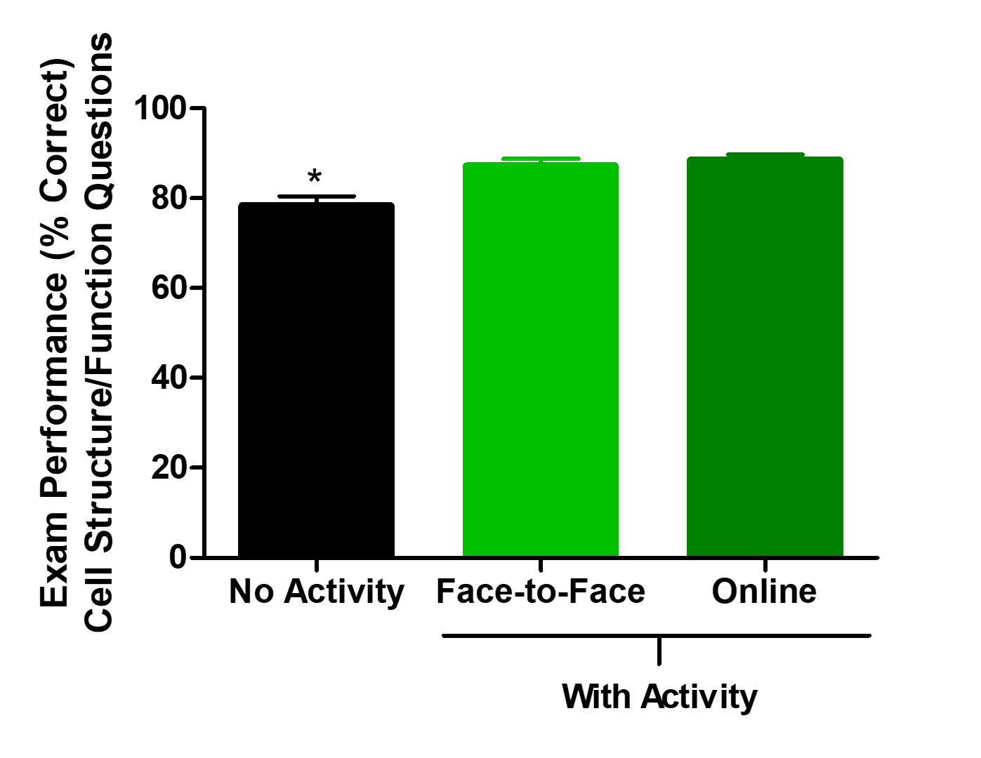
This adaptation of the Cell Engineer/Detective activity is different from the original activity during which students attempt to sketch a cell based on a description of its function. However, I have found it to be equally as effective as the original activity in helping students understand cell structure and function. As indicated in Figure 1, students performed significantly better on the cell structure exam questions when the Cell Engineer/Detective activity was implemented, but there was no difference in performance whether the activity was used as described previously in a face-to-face class or as described here in an online setting. Students also expressed positive views of the activity. When asked which course activity they enjoyed most, one student enrolled in my summer 2019 online course responded, "I enjoyed the activity where we compared the cell cartoons and the pictures of cells we found online! I enjoyed applying what I learned to a real life cell and felt like it was actually showing me that I was understanding what I was learning."

Figure 1. Students performed significantly better on cell structure and function exam questions when they completed the Cell Detective activity (p<0.001). There was no difference in performance between face-to-face and online sections of the course.
SUPPORTING MATERIALS
- S1. Adapting Cell Detective for Online – Reading Quiz
- S2. Adapting Cell Detective for Online – Goblet Cell Images
- S3. Adapting Cell Detective for Online – List of Sample Cell Types
- S4. Adapting Cell Detective for Online – Sample Student Responses
References
- American Association for the Advancement of Science. 2011. Vision and change in undergraduate biology education: A call to action. Washington, DC.
- Alberts B, Hopkin K, Johnson A, Morgan D, Raff M, Roberts K, Walter P. 2019. Essential Cell Biology. New York, NY: W. W. Norton & Company.
- Sestero C, Tinsley H, Ye ZH, Zheng X, Graze R, Kearley M. 2014. Using the Cell Engineer/Detective Approach to Explore Cell Structure and Function. CourseSource. doi: 10.24918/cs.2014.7.
- Fowler S, Roush R, Wise J. 2016. Concepts of Biology. Houston, TX: OpenStax.
- Amoeba Sisters. 2016. Introduction to Cells: The Grand Cell Tour [video]. YouTube. https://www.youtube.com/watch?v=8IlzKri08kk.
Article Files
Login to access supporting documents
Online Adaptation of the Cell Engineer/Detective Lesson(PDF | 221 KB)
S1. Adapting Cell Detective for Online - Reading Quiz.docx(DOCX | 103 KB)
S2. Adapting Cell Detective for Online -Goblet Cell Images.pdf(PDF | 1 MB)
S3. Adapting Cell Detective for Online - Cell Type List.docx(DOCX | 13 KB)
S4. Adapting Cell Detective for Online -Sample Student Responses.docx(DOCX | 15 KB)
- License terms

Comments
Comments
There are no comments on this resource.