How Do Kidneys Make Urine From Blood? Qualitative and Quantitative Approaches to Filtration, Secretion, Reabsorption, and Excretion
Editor: Justin Shaffer
Published online:
Abstract
The function of the kidneys is to help maintain a constant internal environment (homeostasis) by regulating the volume and chemical composition of the blood. This regulation occurs via three fundamental processes: filtration, secretion, and reabsorption. Because these three processes all concern transfers between the blood and the pre-urine, inexperienced biology students frequently confuse them with each other and with the related process of excretion. Such confusion impairs understanding of the kidney’s regulatory functions. For instance, the effects of H+ secretion and HCO3- reabsorption on plasma pH can only be predicted if one knows that secretion entails removal from the blood while reabsorption entails addition to the blood. The enclosed three-part lesson teaches these processes through the use of multiple related examples with clinical relevance. In Module A (“Simple Math”), students define the direction of transfer (blood to pre-urine or pre-urine to blood) for each process, create a simple equation to show how excretion rate depends on these three processes, and solve the equation for missing values. In Module B (“Simple Graphs”), students show qualitatively how the three processes affect the composition of the pre-urine and (by implication) the blood. In Module C (“GFR”), students examine the relationship between glomerular filtration rate (GFR) and plasma levels of solutes like creatinine. By presenting multiple related examples embedded in the framework of Test Question Templates (TQTs), this lesson promotes a solid understanding of filtration, secretion, reabsorption, and excretion that can be applied to any naturally occurring substance or drug.
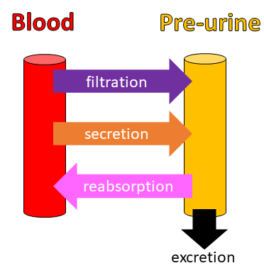
Primary image: Four urinary system processes. This image visually summarizes the four processes covered in this lesson: filtration, secretion, reabsorption, and excretion.
Citation
Crowther GJ. 2021. How Do Kidneys Make Urine From Blood? Qualitative and Quantitative Approaches to Filtration, Secretion, Reabsorption, and Excretion. CourseSource. https://doi.org/10.24918/cs.2021.42
Lesson Learning Goals
- Modules A & B: In the context of renal physiology, students will understand the differences between filtration, secretion, reabsorption, and excretion.
- Modules A & B: Students will understand how the composition of urine is determined by the processes of filtration, secretion, and reabsorption.
- Module C: Students will learn how kidney function can be estimated from plasma levels of a marker solute.
Lesson Learning Objectives
- Module A: Students will be able to calculate a substance’s rate of filtration, secretion, reabsorption, or excretion if given the other three rates.
- Module B: Students will be able to identify a substance’s sites of filtration, secretion, and/or reabsorption from a qualitative graph, or vice versa (i.e., students will be able to draw a qualitative graph to represent filtration, secretion, and/or reabsorption).
- Module B: Students will be able to predict a substance’s persistence in or removal from the body, and its potential as an osmotic diuretic, based on its transport in the nephron and collecting duct.
- Module C: Students will be able to make predictions about glomerular filtration rate (GFR) from plasma concentrations of a marker solute, or vice versa (i.e., students will be able to make predictions about plasma concentrations of a marker solute from GFR).
Article Context
Course
Article Type
Course Level
Bloom's Cognitive Level
Vision and Change Core Competencies
Vision and Change Core Concepts
Class Type
Class Size
Audience
Lesson Length
Pedagogical Approaches
Principles of How People Learn
Assessment Type
Introduction
The process by which kidneys generate urine from blood involves three processes: filtration from glomeruli into glomerular capsules (Bowman’s capsules), secretion into the lumen of nephron tubules and collecting ducts, and reabsorption from the lumen of nephron tubules and collecting ducts into peritubular capillaries. Collectively, these three processes are responsible for both the excretion of metabolic wastes and the regulation of extracellular fluid composition and blood pressure; as such, they deserve the careful attention of physiology students (1). However, undergraduates often fail to distinguish these three processes from each other and from excretion, the overall process to which they contribute (2). The present lesson helps students apply these terms to multiple examples in different contexts, thus clarifying the fundamental differences between filtration, secretion, reabsorption, and excretion (Figure 1).

This lesson employs two complementary strategies for helping students learn biology while honing their quantitative reasoning skills. The first strategy, employed in Modules A (“Simple Math”) and C (“GFR”), is the use of mathematical models to present biological relationships with maximal clarity. Equations can often summarize biological phenomena more precisely than purely verbal descriptions (3; see also Fox 2012). For example, in Module A, students discover the following equation:
Filtration Rate + Secretion Rate – Reabsorption Rate = Excretion Rate
This equation, though simple, clarifies that, for any given substance, increasing the filtration and/or secretion rate will increase the excretion rate, while increasing the reabsorption rate will decrease the excretion rate. Module C employs a different, more complex equation that helps students understand a particular inverse relationship: the higher the plasma concentration of certain solutes such as creatinine, the lower the estimated glomerular filtration rate (GFR), a key indicator of kidney health.
The second strategy, employed in Module B (“Simple Graphs”), is the use of qualitative graphs, i.e., graphs without numerical values. Compared with mathematical equations and quantitative graphs, qualitative graphs have the advantage of focusing attention on general trends, rather than exact values (4,5). Our main concern in Module B is whether the filtered load of a given substance (in units of mass per unit time, e.g., mg/min) is increasing or decreasing in each segment of the nephron/collecting duct, making qualitative graphs an appropriate tool to use.
The biology education literature offers many fine qualitative and quantitative approaches to renal physiology. Previous articles address such important concerns as how to apply general physiology concepts (e.g., Starling forces, cell membrane transport) to the kidneys (6), how to teach functions (e.g., regulation of plasma pH and blood pressure) that are shared by the urinary system and other organ systems (7,8), how to explain notoriously challenging kidney subtopics (9) such as renal clearance (10,11), and how to simulate kidney functions in laboratory exercises (12,13).
The present lesson arguably complements previous work in two respects. First, this lesson is intended for students who are relatively inexperienced in renal physiology (and who therefore may fail to distinguish secretion from excretion, for example), whereas most similar articles concern more advanced students. Second, this lesson explicitly connects the contents of lesson itself to possible future exam questions, thus helping students transfer knowledge from the lesson itself to novel future contexts (14).
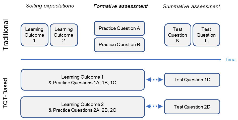
Here our framework for promoting knowledge transfer is that of Test Question Templates. TQTs have been described in detail (15); in brief, a TQT directly bundles a learning outcome (LO) with multiple specific examples of how that LO might be assessed. While most courses come with LOs and practice questions, the links between them are often unclear to students, and links between practice questions and actual test questions are often murky as well. By showing students, as clearly as possible, the relationships between LOs, practice questions, and actual test questions (Figure 2), TQTs should help students study more efficiently with greater motivation and less anxiety.

In the absence of a TQT-like framework, students might prepare for a test on a previous class lesson by rereading the lesson’s questions and answers; however, this approach would be suboptimal, in part because the test’s questions will likely differ (in unspecified ways) from the lesson’s questions. In contrast, if the lesson includes TQTs (as the present lesson does), and if the instructor commits to writing test questions based directly on those TQTs, students can use the TQTs to generate additional new practice questions that are well-aligned to the test, thus reinforcing and extending their understanding of the lesson’s material.
Intended Audience
Introductory physiology students such as sophomore pre-nursing and pre-health sciences students.
Required Learning Time
This lesson is modular, so individual modules (Module A, Module C) can be completed in as little as 10-20 minutes. Altogether, the three modules take 100 to 200 minutes, depending on the details of implementation (see Table 1).
Table 1. Lesson Plan Timeline for Qualitative and Quantitative Approaches to Filtration, Secretion, Reabsorption, and Excretion
| Activity | Description | Estimated Time |
|---|---|---|
| Students review background info | Students learn or review the 3 areas listed under Prerequisite Student Knowledge. This could be done in class or outside of class (via video lectures, textbook readings, etc.). | 30-60 minutes |
| Students do Module A, “Simple Math” (Slides 2-3) | Students learn the locations and directions of filtration, secretion, and reabsorption, and use an equation to relate these to excretion. This could be done in class or as homework. | 10-20 minutes |
| Instructor introduces Module B, “Simple Graphs” (Slides 4-12) | Module B is built around a particular graph format (Figure 1) that will not be familiar to students, so the instructor should introduce the format and demonstrate it for at least one example solute (in class, or as a video lecture). | 5-10 minutes |
| Students do Module B, “Simple Graphs” (Slides 4-12) | Students gain practice translating verbal information about filtration, secretion and reabsorption into qualitative graphs. This module is ideal for small-group work. Each small group can work on the entire worksheet, or a “jigsaw” format can be used. | 30-50 minutes |
| Students do Module C, “GFR” (Slides 13-15) | Students study the inverse relationship between plasma solute concentrations and GFR. Like Module B, this module is challenging enough for most introductory biology students to benefit from working in groups. | 10-20 minutes |
| Students create additional TQT-based examples | Optional: Students can use class time to create additional example questions for an assigned TQT (TQT A, B1, B2, or C – see S1. Urine from Blood – PowerPoint Worksheet), and to quiz each other on these examples. | 10-30 minutes |
Prerequisite Student Knowledge
This lesson assumes that students already have some basic knowledge in the following three areas: (i) the anatomy of nephrons and corresponding blood vessels (afferent and efferent arterioles, glomeruli, peritubular capillaries); (ii) the general functions of the urinary system (maintaining a constant internal environment by regulating the volume and chemical composition of the blood); and (iii) working definitions of filtration, secretion, reabsorption, and excretion in the context of the urinary system. This background could be covered in a prior class period or in a pre-lecture homework assignment.
Prerequisite Teacher Knowledge
This lesson focuses on careful definitions of terms (e.g., distinguishing between reabsorption and secretion), so teachers should review the key terms and decide which distinctions are most important for their purposes. For example, the term “secretion” can broadly refer to the movement of plasma solutes into the lumen of the nephron or collecting duct, but its more precise meaning, in the context of kidney function, is the release of a substance from a renal tubule (or collecting-duct epithelium) cell into the lumen. This detail may or may not be worth noting, depending on one’s audience, goals, and time available. Similarly, it may be tempting to speak broadly of nephrons and nephron tubules, yet, technically, the PowerPoint worksheet goes beyond these structures (collecting ducts are not considered parts of nephrons, and renal corpuscles are not considered parts of nephron tubules). Again, these distinctions may or may not be worth highlighting.
In addition, teachers should make sure that they are familiar with the estimation of glomerular filtration rates (GFR) from plasma levels of solutes like creatinine, inulin, and iothalamate (Slides 13-15 of Supporting File S1. Urine from Blood – PowerPoint Worksheet) (16). Since equations for estimating GFR often include a “race factor” (as shown in Slide 13), teachers should also acquaint themselves with the history and context of such factors (17).
Scientific Teaching Themes
Active Learning
This lesson is essentially a three-part PowerPoint worksheet that students can do individually or (preferably) in groups. Answers should initially be withheld so that students are encouraged to come up with their own answers. Module A is the most straightforward section and could potentially be completed as pre-lesson homework. Since Module B includes several similar but independent examples, it could be covered as a “jigsaw” (18), an active-learning activity in which each student becomes an expert on one example and teaches that example to their peers while learning about the other examples from their peers.
Assessment
This lesson includes four TQTs (15), which provide opportunities for formative assessment while also foreshadowing subsequent summative assessment. Having an explicit template for possible test questions empowers students to keep practicing a given type of problem – creating their own additional examples, if desired – until they are satisfied with their understanding. TQT examples are presented here as short-answer questions to emphasize scientific reasoning, though they can easily be rewritten as multiple-choice questions if desired.
Inclusive Teaching
In presenting strategies for a specific content area (renal physiology), the present lesson does not directly promote general inclusive teaching practices such as instructor self-reflection, instructor empathy for students, and a positive classroom climate (19). However, the lesson does try to promote two other aspects of inclusive teaching: making space for students’ voices and making expectations as clear as possible.
Modules B and C of the lesson are well-suited for group discussions, in part because they include problems too complex for some students to solve individually. Group work can potentially lead to meaningful interactions among group members (20), which might increase students’ sense of belonging in the classroom (21). A variation of Module C (noted in the “Possible Modifications” subsection of the Teaching Discussion below) offers additional discussion opportunities regarding the “race coefficient” in equations for estimating GFR, which may disadvantage Black kidney failure patients relative to non-Black patients.
The lesson might also be considered inclusive in the sense that TQTs potentially provide students with transparent alignment of learning activities and test questions (15). This transparency of expectations should be especially helpful to students who are unfamiliar or uncomfortable with high-stakes testing, students who lack test-savvy study partners, and students for whom reading and writing in English is difficult. Thus, the transparency of TQTs should translate into better inclusion of students facing such challenges.
Lesson Plan
The flow of this lesson, summarized in Table 1, closely follows the PowerPoint worksheet included here as Supporting File S1. Urine from Blood – PowerPoint Worksheet. The worksheet is in the format of a PowerPoint file so fellow instructors can easily edit the arrangement of text and figures, if desired. The PowerPoint worksheet is divided into three parts: Module A: Simple Math (Slides 2-3), Module B: Simple Graphs (Slides 4-12), and Module C: GFR (Slides 13-15). Modules A and B cover the urinary processes of filtration, secretion, reabsorption, and excretion (Figure 2), while Module C focuses on filtration. Module C also differs from the other modules in focusing on the filtration of plasma fluid as a whole (water plus solutes), while Modules A and B cover the filtration of individual solutes.
The PowerPoint worksheet is fairly verbose, and thus may not require much additional explanation; nevertheless, some instructor tips are provided in Table 2. Perhaps the most unconventional aspect of the lesson is the format of the graphs in Module B (Figure 3). Here, the X axis represents the distance that a solute has traveled through a nephron/collecting duct (or parallel blood vessels), while the Y axis indicates the relative amount of the solute passing through the nephron/collecting duct lumen. (The Y axis does not represent a solute concentration per se because concentrations depend on the volume of the solvent, water, which changes with distance along the nephron/collecting ducts due to reabsorption, making inferences about concentrations less straightforward.) The slope of the line at any given point along the X axis indicates whether the solute is being secreted (positive slope), reabsorbed (negative slope), or neither (0 slope), as shown in the left panel of Figure 3.
Table 2. Tips for Instructors on Using Supporting File S1. Urine from Blood – PowerPoint Worksheet.
| Worksheet Section | Notes for Instructors |
|---|---|
| General |
|
| Module A: Simple Math |
|
| Module B: Simple Graphs |
|
| Module C: GFR |
|

Each of the three modules concludes with 1-2 TQTs (15). In brief, a TQT offers a LO (formatted as an input-output statement: given an input, students produce a corresponding output) paired with specific examples of how that LO could be assessed. TQTs thus help students see how specific problems relate to broader patterns of problem-solving, and thus how to solve novel problems that fit a familiar pattern. This important aim – the development of transferable knowledge via practice on multiple related examples – should be explicitly discussed with students, who might otherwise see the numerous examples as onerous. However, students are likely to embrace TQTs, in my experience, if the instructor commits to writing test questions based directly upon them, which assures students that their TQT practice will be highly relevant to the test (15).
For any given TQT, students may vary widely in the number of examples needed to achieve mastery. Instructors can potentially address this heterogeneity by having quick learners coach their peers, which should yield benefits for tutors and tutees alike (22). Such peer-to-peer coaching might include the creation of additional examples beyond those offered in the PowerPoint worksheet, as suggested by the last part of each TQT: “make up an example and ask your classmates!”
Teaching Discussion
This lesson has evolved gradually over several years. As one might surmise, I initially created it in response to my students’ perennial confusion about the meanings of filtration, secretion, reabsorption, and excretion. Since recitations of definitions did not seem adequate for most students, I figured that if they applied the terms to multiple examples with clear right and wrong answers, they would get the needed practice, and might also think metacognitively (23) about their progress.
The first version of the lesson (summer 2016) consisted of the Module B slides as they existed at the time. The lesson was a success in the sense that it helped students realize the extent of their confusion; they had lots of trouble answering the questions, prompting further discussion and practice. That first version had several significant limitations, as summarized in Table 3. Perhaps the biggest limitation was that, as a graphing exercise, it was of limited practical interest to most students because it did not yield much insight into practical health issues. However, the current version of the lesson seems to connect more successfully to students’ interests. In particular, many students seem to enjoy figuring out which of two chemically similar drugs will stay in the body longer based on Module-B graphs like the one shown in the right panel of Figure 3. Many of them also seem interested in GFR estimates as indicators of kidney health and transplant eligibility, as covered in Module C.
Table 3. Evolution of a Lesson on Filtration, Secretion, Reabsorption, and Excretion
| Pedagogical Problem | Pedagogical Adjustment [time of change] |
|---|---|
| Many introductory physiology students do not correctly distinguish between filtration, secretion, reabsorption, and excretion. | An initial version of Module B is created. [Summer 2016] |
| The lesson does not clearly connect a substance’s concentration in the collecting duct to its excretion rate. | Questions on sustances’ persistence in the body are added to Module B. [Spring 2019] |
| Some students struggle to transfer their understanding of Module B to additional new scenarios. | Test Question Templates are added to Module B. [Winter 2020] |
| Despite covering filtration, the lesson does not cover common clinical measurements and estimates related to filtration (e.g., plasma [creatinine], eGFR, clearance). | Module C is created to cover plasma [creatinine], eGFR, and clearance. [Fall 2020] |
| Student are confused by the concept of clearance (C), a virtual volume of blood per unit time from which a substance has been completely eliminated. | Coverage of clearance (C) is removed from Module C. [Winter 2021] |
| The lesson dives straight into complicated graphs of filtration, secretion, and reabsorption. | Module A is created as a light warm-up to Module B. [Winter 2021] |
| The lesson does not cover diuretics, which are clinically important. | Questions on osmotic diuresis are added to Module B. [Summer 2021] |
Possible modifications
Although this lesson’s three modules complement each other, they are modular, meaning that each module can be used alone or in combination with one or both other modules, in any order (though Module A is easiest, and often a good starting point). Therefore, consistent with the principle of backwards design (24), instructors interested in this lesson should consider which of the Learning Goals and Learning Objectives they have time for and want their students to accomplish, then choose among the modules accordingly.
The exchange of substances between the blood and the pre-urine is, among other things, an illustration of the physiological core concept of mass balance (25). Mass balance tells us that if a substance is accumulating in the pre-urine, it must be disappearing from the blood, and vice versa. However, the graphs in Module B do not highlight this conservation of mass, since they only show a solute’s entry into or exit from the pre-urine without showing the corresponding (inverse) changes in the blood. Therefore, as an optional extension to Module B, one could challenge students to illustrate a scenario (e.g., glucose in Slide 7) in a way that emphasizes mass balance, either as a graph or in some other visual form.
As written, the present lesson is intended for courses in human physiology, rather than comparative animal physiology. Nephrons are structurally and functionally similar among vertebrates, except that only birds and mammals have nephron loops (loops of Henle), allowing them to produce concentrated urine (26). The present lesson thus could be adapted for comparative animal physiology courses, and could be useful in that context, but is not set up to explain the unique osmoconcentration abilities of birds and mammals, which may be covered better by other approaches (27).
Advanced students could use Module C (GFR) as a jumping-off point for further learning about renal clearance rate (C), another clinically relevant parameter. C is equal to GFR for substances (e.g., inulin) that are neither secreted nor reabsorbed, but is different otherwise. Since C is a virtual volume of blood processed per unit time, it is nonintuitive for most introductory biology students (10), though accessible to most upper-level undergraduates, graduate students, and medical students.
Finally, the GFR equation used in Module C could provide a starting point for a bioethics lesson or discussion on the meaning of race in a biomedical context and when (if ever) race should be factored into medical consultations and treatments (28). The use of a “race coefficient” in estimating GFR is not merely hypothetical; equations similar to the one in Module C continue to be used in clinics, and ongoing research suggests that continued use of such coefficients may propagate and amplify other health-care inequities (17). In my experience, such discussions would likely touch upon the following issues: whether a binary racial choice (Black or non-Black) makes sense here, whether the equation’s binary approach to biological sex (male or female) is OK or problematic, who should decide which race and which sex are entered (patient or care team or both), and how care providers might maintain sensitivity and scientific validity when following protocols involving rigid demographic categories.
Conclusion
Years before the concept of scientific teaching was formalized (29), renal physiologist Linda Costanzo (30) informally presaged some of its features with the following exhortation:
Our job is to teach with integrity, which means to give the students everything we have. By ‘‘everything we have,’’ I do not mean delivering the material on a silver platter in lecture. I mean bombarding the students with as many examples as we can, reinforcing principles as often as we can, and endeavoring to reach every single student, weak and strong.
In a recent review of cognitive psychology literature, Kaminske et al. (14) found support for Costanzo’s use of multiple examples. In particular, lessons providing multiple examples with different “surface features” were found to help students learn to transfer their knowledge to unfamiliar contexts.
The present lesson on renal physiology adopts and refines the practice of offering multiple superficially different but related examples. Here, the examples are organized into frameworks (TQTs) that help students recognize and act upon the examples’ underlying commonalities. The explicit linking of specific examples to more general LOs should help students see biology as something more than a collection of facts and should facilitate their development of knowledge that is truly transferable.
SUPPORTING MATERIALS
S1. Urine from Blood – PowerPoint Worksheet
Acknowledgments
I thank Dilan Evans and Lekelia Jenkins of Arizona State University, Leila Zelnick of the University of Washington, Joel Michael of Rush Medical College (emeritus), and an anonymous reviewer for their input on the planning, writing, and revision of this article.
References
- Medler S, Harrington F. 2013. Measuring dynamic kidney function in an undergraduate physiology laboratory. Adv. Physiol. Educ. 37(4):384-391. doi: 10.1152/advan.00057.2013.
- Dirks-Naylor AJ. 2016. An active learning exercise to facilitate understanding of nephron function: anatomy and physiology of renal transporters. Adv. Physiol. Educ. 40(4):469-471. doi: 10.1152/advan.00111.2016.
- Horton RM, Leonard WH. 2013. Some applications of mathematics for the biology classroom. Am. Biol. Teach 75(4):281-284. doi: 10.1525/abt.2013.75.4.10.
- Summers RL, Woodward LH, Sanders DY, Hall JE. 1996. Graphic analysis for the study of metabolic states. Adv. Physiol. Educ. 15:S81-87. doi: 10.1152/advances.1996.270.6.S81.
- Matuk C, Zhang J, Uk I, Linn MC. 2019. Qualitative graphing in an authentic inquiry context: How construction and critique help middle school students to reason about cancer. Journal of Research in Science Teaching 56(7):905-936. doi: 10.1002/tea.21533.
- Roosa KA. 2021. Engaging undergraduates in mechanisms of tubular reabsorption and secretion in the mammalian kidney. CourseSource 8:4. doi: 10.24918/cs.2021.4.
- Longmuir KJ. 2014. Interactive computer-assisted instruction in acid-base physiology for mobile computer platforms. Adv. Physiol. Educ. 38(1):34-41. doi: 10.1152/advan.00083.2013.
- Waghmare LS, Srivastava TK. 2016. Conceptualizing physiology of arterial blood pressure regulation through the logic model. Adv. Physiol. Educ. 40(4):477-479. doi: 10.1152/advan.00074.2016.
- Vander AJ. 1998. Some difficult topics to teach (and not to teach) in renal physiology. Adv. Physiol. Educ. 20:S148-156. doi: 10.1152/advances.1998.275.6.S148.
- Hull K. 2016. Renal clearance: using an interactive activity to visualize a tricky concept. Adv. Physiol. Educ. 40(4):458-461. doi: 10.1152/advan.00059.2016.
- Baptista V. 2020. A didactic approach to renal clearance. HAPS Educator 24(3):54-60. doi: 10.21692/haps.2020.022.
- Whiting CC. 2015. Human Anatomy & Physiology Laboratory Manual: Making Connections. Boston, MA: Pearson.
- Motz VA, Suniga RG, Connour JR. 2018. Inexpensive hands-on activities to reinforce basic physiological principles: Details of a soda bottle nephron model. HAPS Educator 22(2):159-164. doi: 10.21692/haps.2018.017.
- Kaminske AN, Kuepper-Tetzel CE, Nebel CL, Sumeracki MA, Ryan SP. 2020. Transfer: A review for biology and the life sciences. CBE Life Sci. Educ. 19(3): es9. doi: 10.1187/cbe.19-11-0227.
- Crowther GJ, Wiggins BL, Jenkins LD. 2020. Testing in the age of active learning: Test Question Templates help to align activities and assessments. HAPS Educ. 24(1):74-81. doi: 10.21692/haps.2020.006.
- Pottel H, Hoste L, Dubourg L, Ebert N, Schaeffner E, Eriksen BO, Melsom T, Lamb EJ, Rule AD, Turner ST, Glassock RJ. 2016. An estimated glomerular filtration rate equation for the full age spectrum. Nephrol. Dial. Transplant. 31(5):798-806. doi: 10.1093/ndt/gfv454.
- Boulware LE, Purnell TS, Mohottige D. 2021. Systemic kidney transplant inequities for black individuals: Examining the contribution of racialized kidney function estimating equations. JAMA Netw. Open 4(1):e2034630. doi: 10.1001/jamanetworkopen.2020.34630.
- Colosi JC, Zales CR. 1998. Jigsaw cooperative learning improves biology lab courses. Bioscience 48(2):118-124. doi: 10.2307/1313137.
- Dewsbury B, Brame CJ. 2019. Inclusive teaching. CBE Life Sci. Educ. 18(2):fe2. doi: 10.1187/cbe.19-01-0021.
- Wilson KJ, Brickman P, Brame CJ. 2018. Group work. CBE Life Sci. Educ. 17(1):fe1. doi: 10.1187/cbe.17-12-0258.
- Wilton M, Gonzalez-Niño E, McPartlan P, Terner Z, Christoffersen RE, Rothman JH. 2019. Improving academic performance, belonging, and retention through increasing structure of an introductory biology course. CBE Life Sci. Educ. 18(4):ar53. doi: 10.1187/cbe.18-08-0155.
- Chrispeels HE, Klosterman ML, Martin JB, Lundy SR, Watkins JM, Gibson CL, Muday GK. 2014. Undergraduates achieve learning gains in plant genetics through peer teaching of secondary students. CBE Life Sci. Educ. 13(4):641-652. doi: 10.1187/cbe.14-01-0007.
- Tanner KD. 2012. Promoting student metacognition. CBE Life Sci. Educ. 11(2):113-120. doi: 10.1187/cbe.12-03-0033.
- Wiggins G, McTighe J. 2005. Understanding by Design [2nd edition]. Alexandria, VA: Association for Supervision and Curriculum Development (ASCD).
- Michael J, Modell H. 2021. Validating the core concept of “mass balance.” Adv. Physiol. Educ. 45: 276-280. doi: 10.1152/advan.00235.2020.
- Sherwood L, Klandorf H, Yancey PH. 2013. Animal Physiology: From Genes to Organisms. Belmont, CA: Brooks/Cole.
- Katz SA. 1998. Some teaching tips on the mechanisms of urinary concentration and dilution: countercurrent multiplication be damned. Adv. Physiol. Educ. 20:S195-S205. doi: 10.1152/advances.1998.275.6.S195.
- Nieblas-Bedolla E, Christophers B, Nkinsi NT, Schumann PD, Stein, E. 2020. Changing how race is portrayed in medical education: recommendations from medical students. Acad. Med. 95(12):1802-1806. doi: 10.1097/ACM.0000000000003496.
- Handelsman J, Miller S, Pfund C. 2007. Scientific Teaching. New York, NY: W. H. Freeman and Company.
- Costanzo LS. 1998. Good teaching and good testing: Examples from renal physiology. Adv. Physiol. Educ. 20:S217-220. doi: 10.1152/advances.1998.275.6.S217.
Article Files
Login to access supporting documents
Crowther-How do kidneys make urine from blood.pdf(PDF | 393 KB)
S1.Urine from Blood - PowerPoint Worksheet.pptx(PPTX | 2 MB)
- License terms

Comments
Comments
There are no comments on this resource.