Enhancing the Microscopy Skills of Non-Science Majors and Nursing Microbiology Students: Promoting the Practice of Observing Multiple Fields of View Using Blood Smear Slides
Editor: Heather Townsend
Published online:
Abstract
One of the challenges in teaching microscopy is having students scan multiple fields of view at high power magnification. Many times, students will feel this unnecessary, especially when presented with slides that show only one structure or a monoculture of cells. This communication presents a simple microscopy activity to engage students in the importance of examining several fields of view when using the microscope. Students are challenged with determining whether an “unknown” blood smear slide is indicative of normal blood or a blood disorder. The disorders the activity examines include sickle cell anemia, leukemia, thrombocytosis and a bloodstream infection. All slides can be purchased from science education supply companies. Students must properly focus on commercially available blood slides and examine several fields of view in order to reach the most reasonable diagnosis. This lesson has been used to engage both non-science majors taking a laboratory-based science class as well as nursing/allied health microbiology students and simulates real-life scenarios in diagnosis.
Primary Image: Blood smear showing a bloodstream infection. (Photo: B.M. Forster)
Citation
Forster BM, Pacitti AF. 2023. Enhancing the Microscopy Skills of Non-Science Majors and Nursing Microbiology Students: Promoting the Practice of Observing Multiple Fields of View Using Blood Smear Slides. CourseSource 10. https://doi.org/10.24918/cs.2023.32
Article Context
Course
Article Type
Course Level
Bloom's Cognitive Level
Vision and Change Core Competencies
Vision and Change Core Concepts
Class Type
Class Size
Audience
Lesson Length
Principles of How People Learn
Assessment Type
Introduction
Microscopy is an integral part of the biology laboratory. It is a skill that has been identified in ASM’s Curriculum Guidelines for Undergraduate Microbiology Education (1), Microbiology in Nursing and Allied Health (MINAH) Undergraduate Curriculum Guidelines (2) and the Condensed MINAH laboratory curriculum (3). When first introducing the microscope, instructors often utilize slides that either have one structure on it or a monoculture of cells. Instructors use these types of slides to have students practice their focusing skills. We have observed that students typically focus on the first field of view only. A field of view is the area visible when looking through a microscope’s eyepiece. It doesn’t naturally occur to students to scan around and get a view of the slide as a whole instead of the one spot they focused on. It is also possible instructors forget to instruct or remind students to do so.
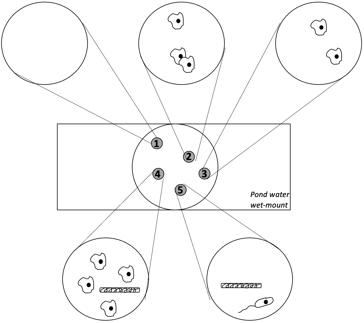
Students learning how to use the microscope should be taught to scan multiple fields of view (Figure 1). Students may miss different cellular structures if they only look at one field of view. To encourage students to scan multiple fields of view while using a microscope, we have developed a simple laboratory activity using prepared blood slides. This activity can be integrated into any introductory microscopy lab exercise. It is an activity that can be completed in approximately a half hour. This activity supports the Microbiology Learning Framework of Cell Structure and Function (1), especially the question of “How have the structure and function of microorganisms been revealed by the use of microscopy?” Without proper knowledge of the microscope, students cannot answer this question fully.

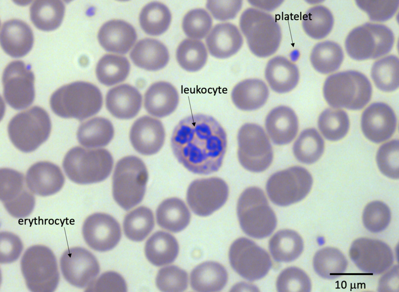
Blood is liquid connective tissue that the body uses for transport, immunity, salinity and temperature regulation (4). Erythrocytes (red blood cells) carry gases throughout the body using hemoglobin. Leukocytes (white blood cells) are the cellular components of the immune system. Platelets are membrane bound cellular fragments important for clotting. Blood smear slides are typically stained with either Giesma or Wright’s stain to allow for differentiation of blood features (Figure 2). This makes identifying the components of blood rather easy, even for novice microscopists. Students that take data from multiple fields of view can come to a conclusion on the overall health of a simulated patient based on the shape of cells, number of cells and platelet counts. By taking information from multiple, separate fields of view, students may diagnose a variety of blood-borne diseases (5, 6).

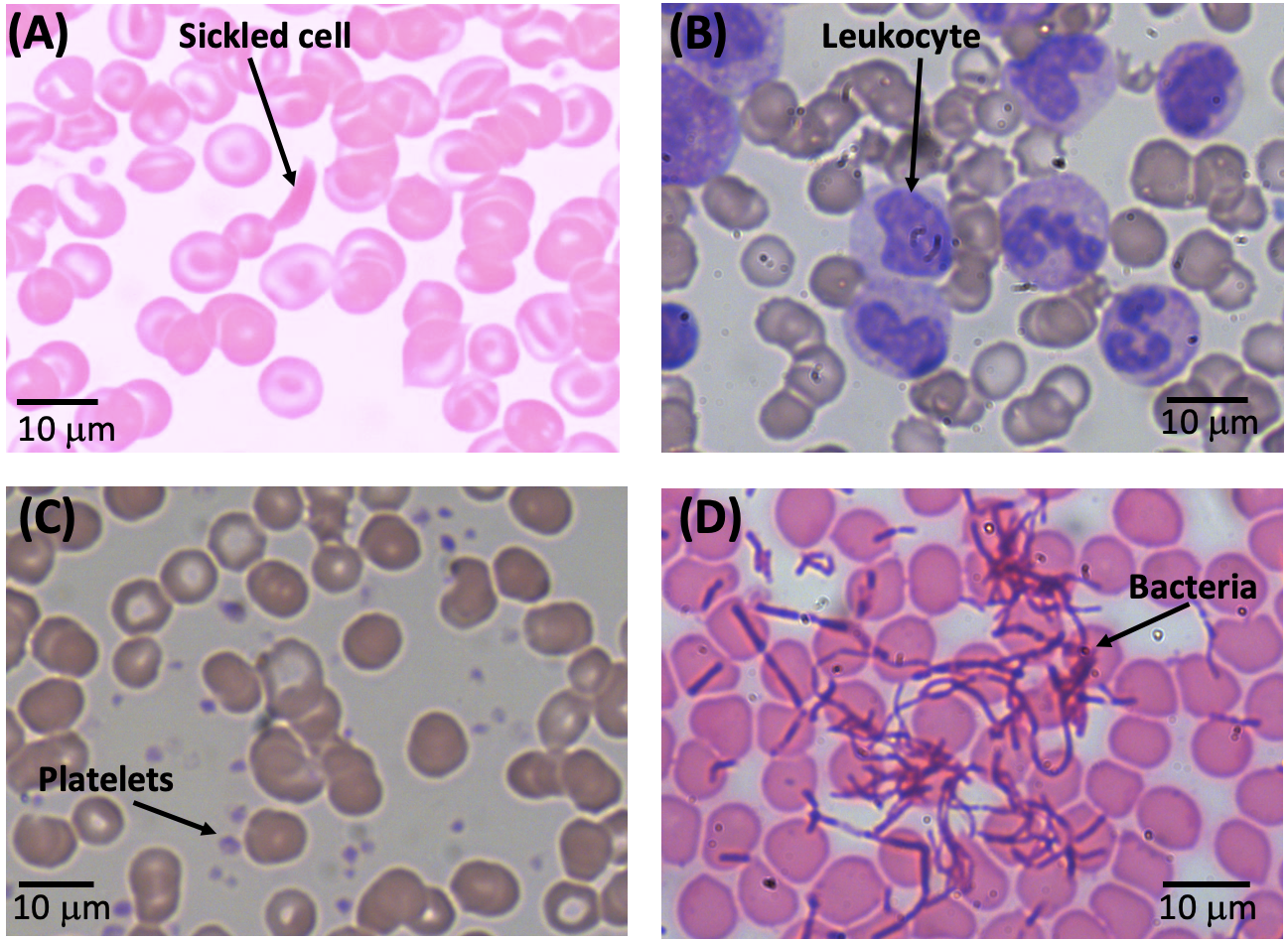
This activity focuses on four blood disorders (Figure 3). Sickle cell anemia (Figure 3A) occurs when erythrocytes have a sickled shape rather than the normal biconcave disc shape (7). This change is due to a mutation in hemoglobin. Individuals can either be homozygous or heterozygous for sickle cell disease. Heterozygotes will express both normal and abnormal hemoglobin, resulting in a mixture of both properly shaped and sickled shape cells. Sickle cell disease affects 100,000 people in the United States (8). Leukemia (Figure 3B) is cancer of leukocytes that often present as the presence of excessive immature cells (9). This results in an increased overall count of leukocytes in the blood and can be caused by numerous factors. One such factor is a chromosomal translocation between chromosomes 9 and 22 (10). It is estimated there will be over 59,000 cases of leukemia in 2023 in the United States (11). Thrombocytosis (Figure 3C) causes an increase in platelet counts (12). Causes of thrombocytosis include a mutation in the thrombopoietin gene and/or infectious disease. One to 2.5 individuals per 100,000 can have thrombocytosis yearly (13). Bacteremia (Figure 3D) is the presence of bacteria in the blood, which in healthy individuals should be sterile. These bloodstream infections can be caused by either a pathogen or can arise from the body’s own microbiome (14).

Examination of multiple fields of view is critical to correctly diagnosing the “patient” with a disease. For example, leukemia may be missed due to the fact that leukocytes may amass in numbers in one area of the blood. Therefore, by viewing a sample of multiple, random sites on the blood slide, large groupings of leukocytes are more likely to be seen. This procedure is common practice in diagnostic laboratories and is essential for effective community healthcare. In this lesson, students assume the role of diagnosticians. Just like professional diagnostic labs, students must use multiple fields of view to determine whether a fictitious patient has a blood disorder. In the next section, we describe how the lesson can be implemented.
Procedure
The materials required for this activity are a microscope capable of at least 400x total magnification and prepared blood smear slides. This includes normal human blood, sickle cell anemia, leukemia, thrombocytosis and bacteremia slides. These slides can be obtained from science lab supply companies including Carolina Biological, Wards Science or Flinn Scientific. Prior to giving slides to the students, instructors should rename the slides “Patient A,” “Patient B,” etc., to make it a mystery and cover up the diagnosis. A student handout instructors can distribute to students can be found in the supporting materials (Supporting File S1).
Students should know how to properly focus on a slide at both low and high power. Prior to starting this activity, instructors should review the components of blood. Instructors can use Figure 2 to aid them in introducing this topic. Instructors should also show students prior to beginning the activity what bacteria look like, focusing on spherical (coccus) and rod-shaped (bacillus) bacteria. To begin the activity, each student (or student group) receives a normal blood smear slide. Students should become proficient in recognizing normal blood parameters first. Students should begin by focusing on the slide at resolving low power and then work their way up to high power (400x or 1000x magnification).
Students should obtain a patient slide and begin focusing on the slide at low power and then move to high power. Once the slide is in focus, students record their observations for five separate fields of view. Students count the number of different cell types and platelets present in the different fields to assess their relative abundance. A sample blank data table can be found in the student handout in the supporting materials (Supporting File S1). After the students have recorded their observations, the instructor distributes the blood reference table found in the supporting materials (Supporting File S2). Students then compare their observations to this information and then try to reach a conclusion as to what blood disorder (if any) the patient has. It is important to note that using the sickle cell blood smear will probably be the hardest to interpret. As seen in Figure 3, not all the erythrocytes on the slide will be sickled, especially if the individual is heterozygous (7).
This activity can be completed either in a lab (approximately 30–45 minutes) or as part of a take home lab (for online classes) where students are asked to purchase a microscope and prepared slides as part of a kit. Alternatively, for online labs that do not have a kit, the instructor can post images of several blood smears, asking the student to identify the structures, complete the worksheet (see Supporting File S1) and make the appropriate diagnoses. Figure 3 shows representative images of each blood disorder.
Active Learning and Assessment
This activity promotes active learning as students must use the microscope (or examine micrograph images if done virtually) to identify whether a blood smear is indicative of normal blood or a blood disorder. The students have to reach their own conclusions from the microscopy observations and data they collect. Instructors can assign this activity as part of an introductory lab activity on using the microscope. As part of a submitted lab report, students complete the student handout (see Supporting File S1), recording their observations of their assigned blood smear. Students are then asked to identify what blood disorder (if any) their assigned patient has. They must explain how they arrived at their answer, comparing the data they collected to a blood reference table (see Supporting File S2).
Conclusion
This activity demonstrates to students the importance of examining multiple fields of view and the importance of adequate sampling. It is not enough for us to simply teach students how to focus on a slide. We have used this activity in our non-science major lab classes and our MINAH courses. Students in these courses have reported enjoying this activity since it allows them to see a real-world medical application using microscopy. This is consistent with other reports of using disease diagnoses as a means of engaging non-science majors (15). For more advanced classes, this activity can be expanded upon in different ways; students could identify the specific type of leukocytes observed and/or measure the size of the different cells and platelets observed. Students could be given additional blood disorder slides, such as eosinophilia or iron deficiency anemia. For MINAH students, this activity encourages students to develop increased care and due diligence of their responsibility to patients to make best diagnostic opinions for best possible outcomes.
Supporting Materials
-
S1. Enhancing Microscopy Skills – Student Handout
-
S2. Enhancing Microscopy Skills – Blood Reference Table
Acknowledgments
The authors would like to acknowledge Karl Siegert, Joni Baumgarten, Taufiqul Huque, Chris Knob, Steve Levin, Riva Marcus, C. Grier Sellers and Jordan Teisher for their suggestions on the development of this activity and for using this activity in their laboratory classes.
References
- Merkel S, ASM Task Force on Curriculum Guidelines for Undergraduate Microbiology. 2012. The development of curricular guidelines for introductory microbiology that focus on understanding. J Microbiol Biol Educ 13:32–38. doi:10.1128/jmbe.v13i1.363.
- Norman-McKay L, ASM MINAH Undergraduate Curriculum Guidelines Committee. 2018. Microbiology in Nursing and Allied Health (MINAH) Undergraduate Curriculum Guidelines: A call to retain microbiology lecture and laboratory courses in nursing and allied health programs. J Microbiol Biol Educ 19:10.1128/jmbe.v19i1.1524. doi:10.1128/jmbe.v19i1.1524.
- McCall B, Pacitti AF, Forster BM. 2019. A simplified but comprehensive laboratory curriculum for microbiology in nursing and allied health (MINAH) courses. J Microbiol Biol Educ 20:10.1128/jmbe.v20i3.1841. doi:10.1128/jmbe.v20i3.1841.
- Glenn A, Armstrong C. 2019. Physiology of red and white blood cells. Anaesth Intensive Care Med 20:170–174. doi:10.1016/j.mpaic.2019.01.001.
- Deshpande NM, Gite S, Aluvalu R. 2021. A review of microscopic analysis of blood cells for disease detection with AI perspective. PeerJ Comput Sci 7:e460. doi:10.7717/peerj-cs.460.
- Bain BJ. 2005. Diagnosis from the blood smear. N Engl J Med 353:498–507. doi:10.1056/NEJMra043442.
- Williams TN, Thein SL. 2018. Sickle cell anemia and its phenotypes. Annu Rev Genomics Hum Genet 19:113–147. doi:10.1146/annurev-genom-083117-021320.
- Hassell KL. 2010. Population estimates of sickle cell disease in the U.S. Am J Prev Med 38:S512–S521. doi:10.1016/j.amepre.2009.12.022.
- Davis AS, Viera AJ, Mead MD. 2014. Leukemia: An overview for primary care. Am Fam Physician 89:731–738.
- Sampaio MM, Santos MLC, Marques HS, Gonçalves VLS, Araújo GRL, Lopes LW, Apolonio JS, Silva CS, Santos LKS, Cuzzuol BR, Guimarães QES, Santos MN, de Brito BB, da Silva FAF, Oliveira MV, Souza CL, de Melo FF. 2021. Chronic myeloid leukemia-from the Philadelphia chromosome to specific target drugs: A literature review. World J Clin Oncol 12:69–94. doi:10.5306/wjco.v12.i2.69.
- Siegel RL, Miller KD, Wagle NS, Jemal A. Cancer statistics, 2023. CA Cancer J Clin 2023;73:17–48. doi:10.3322/caac.21763.
- Schafer AI. 2002. Thrombocytosis: Too much of a good thing? Trans Am Clin Climatol Assoc 113:68–77.
- Meier B, Burton JH. 2014. Myeloproliferative disorders. Emerg Med Clin 32:597–612. doi:10.1016/j.emc.2014.04.014.
- Taur Y, Pamer EG. 2013. The intestinal microbiota and susceptibility to infection in immunocompromised patients. Curr Opin Infect Dis 26:332–337. doi:10.1097/QCO.0b013e3283630dd3.
- Garcia R, Rahman A, Klein JG. 2015. Engaging non–science majors in biology, one disease at a time. Am Biol Teach 77:178–183. doi:10.1525/abt.2015.77.3.5.
Article Files
Login to access supporting documents
Forster-Pacitti-Enhancing the Microscopy Skills of Non-Science Majors and Nursing Microbiology Students Promoting the Practice of Observing Multiple Fields of View Using Blood Smear Slides.pdf(PDF | 733 KB)
S1. Enhancing Microscopy Skills - Student Handout.docx(DOCX | 24 KB)
S2. Enhancing Microscopy Skills - Blood Reference Table.docx(DOCX | 19 KB)
- License terms

Comments
Comments
There are no comments on this resource.